EN
EN


Clinical and radiographic efficacy of subtalar screw arthroereisis in the treatment of pediatric flexible flatfoot
Clinical and radiographic efficacy of subtalar screw arthroereisis in the treatment of pediatric flexible flatfoot
Farouk Khury, Emile Danto, Rita Taurman, Martin Faschingbauer
Received: 18 November 2025 / Accepted: 11 January 2026
© The Author(s) 2026
Abstract
Background Subtalar arthroereisis has been reported to be an effective treatment technique for flexible flatfoot (FF) in children. Although many devices for this procedure exist, arthroereisis using screws is still globally used. Therefore, this study aims to revisit subtalar screw arthroereisis (SSA) and investigate its outcomes.
Methods We retrospectively reviewed 353 flexible flatfeet in 178 pediatric patients who underwent SSA between 2007 and 2020. Clinical and radiological assessments were conducted pre-implantation and pre-explantation. Radiographic angles were measured to quantify correction. Statistical analyses included chi-squared tests and Student's t-tests to evaluate clinical improvement and the impact of variables on outcomes.
Results The mean patient age at implantation was 11.96 years. 96.31% of feet showed clinical improvement postoperatively. Radiographic analysis demonstrated significant correction in most (83.33%) angular measurements, with the calcaneal pitch showing the strongest effect size. Postoperative complications occurred in 41.08% of FF, predominantly pain, and were mainly (84.13%) resolved with non-surgical treatment. 4.25% required implant revision, which was significantly more frequent in the younger age and female group.
Conclusion SSA for treatment of FF in children showed favorable results regarding improved clinical aspects and radiographic measurements. Nevertheless, an accurate indication for surgical treatment is necessary.
Level of evidence Clinical retrospective research—Level III.
Keywords Flatfoot arthroereisis · Pediatric flexible flatfoot · Subtalar arthroereisis · Subtalar screw arthroereisis · SSA
Introduction
Pediatric flatfoot is a complex three-dimensional deformity of the foot caused by collapse of medial arch, plantar tilt of talus, and eversion of calcaneus. It is one of the most common pediatric skeletal disorders and a frequent cause for clinical orthopedic consultation. Although the current treatment approach for asymptomatic flexible flatfoot (FF) is "watch-and-wait", intervention may be required in cases of pain and limited activity. Treatment varies from non-surgical activities, such as shoe modification, nonsteroidal anti-inflammatory medications (NSAIDs), physiotherapy, and stretching exercises, addressing comorbidities such as obesity, hypotonia, and ligamentous hyperlaxity, to surgical soft tissue procedures, realignment osteotomies, and subtalar joint non-fusion procedures.
Ever since its first description in the literature by Chambers in 1946, subtalar arthroereisis (SA) has been one of the most widely debated minimally invasive procedures for the treatment of symptomatic FF. The controversy is due to the fact that although several studies showed that it can improve pain, deformity, and function in FF, it also carries relatively high rates of complications (notably sinus tarsi pain and implant removal), inconsistent outcomes, poorly defined indications/contraindications (especially age, severity, and whether to use adjunctive procedures), and a lack of high-quality long-term evidence. Despite reports of various SA techniques and implants, the orthopedic principles implemented remain undisputed—correction of the excessive foot pronation, hindfoot valgus, and medial arch height by introducing an implant directly or indirectly into the sinus tarsi. Among these options, the screw remains a widely favored implant due to its simplicity, rigid mechanical reliability, and widespread availability compared to newer bioabsorbable or expandable devices. However, these advantages come with the notable drawback of frequently requiring a second procedure for implant removal.
While several clinical, radiological, kinematical, biomechanical, and pedobarographic reports have been published, the exact clinical and radiological outcomes of pediatric FF treated with subtalar screw arthroereisis (SSA) remain unclear. Therefore, this study, which included a high volume of pediatric patients with FF, was conducted to describe the outcomes following SSA, by answering the following questions: 1. What are the outcomes following SSA? 2. Does SSA lead to a clinical improvement of pediatric FF? 3. Can SSA assist in radiological correction of pediatric FF? 4. Which factors correlate to the development of postoperative complications?
Methods
Cohort
Study design
This study is a retrospective review of pediatric patients treated for FF with the SSA technique in our institution between 2007 and 2020.
The course of care was as follows:
1. Baseline visit: patients were initially seen, clinically and radiologically examined, and diagnosed with FF based on their clinical and radiological evaluation in our ambulatory outpatient clinic. As part of the preoperative assessment, all patients had a documented history of symptoms and underwent standardized non-operative management, including activity modification, supportive footwear, stretching/physiotherapy, and orthotic insoles. In cases where nonoperative treatment yielded no pain relief and no significant radiological improvement within three months, surgical treatment using SSA was then offered after the exhaustion of conservative measures.
2. Implantation: patients had undergone SSA.
3. First post-implantation follow-up: clinical and radiological examination six weeks following implantation.
4. Second post-implantation follow-up: clinical and radiological examination before implant removal.
5. Explantation: patients had undergone removal of screw either due to reaching skeletal maturity or a complication.
6. Post-explantation follow-up: clinical examination six weeks following implant removal.
The mean interval between pre-implantation and pre-explantation radiographs was 49.76 months (range 4 to 135 months).
Postoperative complications were defined as postoperative sensory deficits, pain (following traumatic injury or without traumatic injury), wound healing disorders (defined as delayed healing or dehiscence, excluding surgical site infections), peroneal contracture or spasm, and fractures.
Patient selection criteria and surgical indication
Included in this study were children with remaining skeletal growth, idiopathic symptomatic FF, were not previously operated, had no neurogenic or neuromuscular pathologies including rigid FF, had complete pre- and postoperative clinical and radiological documentations, and had undergone two surgeries: implantation during skeletal growth, and explantation following skeletal growth arrest, of a subtalar arthroereisis screw in our institution. Skeletal growth arrest was assessed using radiographs to evaluate physical closure, with consideration of the patient's chronological age and expected skeletal maturity.
Diagnosis of idiopathic flexible flatfoot was established based on a combination of clinical and radiographic findings. Clinically, patients presented with a collapsed medial longitudinal arch, hindfoot valgus, and forefoot abduction that corrected with a heel raise (Jack's Test). Radiographically, lateral and dorsoplantar weight-bearing radiographs demonstrated characteristic angular deviations including a Meary's angle greater than 4°, a calcaneal pitch less than 20°, a Costa-Bartani angle greater than 125°, a talar declination angle greater than 30°, a talocalcaneal angle often greater than 35°, and a talometatarsal angle often greater than 20°. Surgical intervention with SSA was indicated for symptomatic flexible flatfoot after exhaustion of conservative measures (such as shoe modification, nonsteroidal anti-inflammatory medications, physiotherapy, and stretching exercises) and in cases of pain and limited activity.
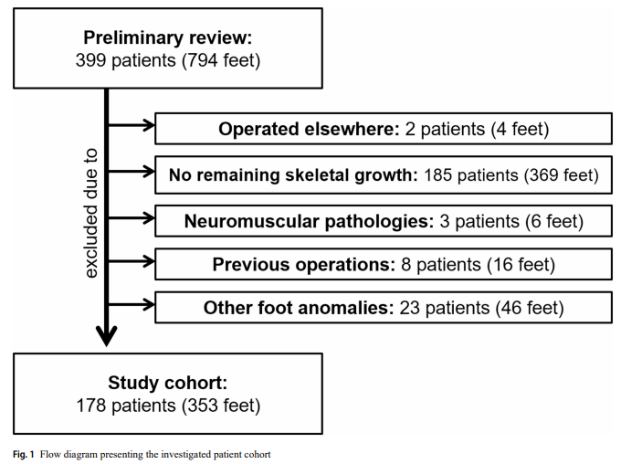
Primary database query yielded the data of 399 potential patients (794 feet) treated with SSA: two patients (four feet) were excluded due to being operated elsewhere, 185 patients (369 feet) were excluded because they had no remaining skeletal growth, reflecting skeletal maturity, three patients (six feet) were excluded due to neuromuscular disorders, eight patients (16 feet) were excluded due to previous operations, and 23 patients (46 feet) were excluded due to other foot anomalies. Following the exclusion process, 178 patients (353 feet) met the inclusion criteria and were eligible for analysis (Fig. 1).
Subtalar screw arthroereisis
Implant
Patients were implanted with either cannulated or non-cannulated stainless steel cancellous screw with available lengths ranging from 25 to 40 mm and diameter of 6 or 6.5 mm.
Operative technique
All surgeries were performed by two experienced attending surgeons in the pediatric orthopedics unit. Information regarding the operative technique is available in supplemental file.
Postoperative care
Immediate postoperative full weight-bearing, as tolerated, was allowed. Skin sutures were removed on postoperative day 14. Sport activities were forbidden for six weeks. Patients were presented for an ambulatory clinical and radiological follow-up six weeks following the surgery. Clinical improvement was established once pain and discomfort were reduced, the excessive foot pronation and hindfoot valgus were corrected, and the medial arch was improved. In the absence of a validated patient-reported outcome instrument, 'clinical improvement' was defined as a composite of subjective symptom relief and objective physical signs assessed by a pediatric foot and ankle specialist with over 20 years of experience. Specifically, improvement was identified by: (1) restoration of the medial longitudinal arch, (2) correction of hindfoot valgus to a neutral or slight varus position, and (3) a neutral forefoot without abduction.
Radiographs
Protocols
Information available in supplemental file.
Angular measurements
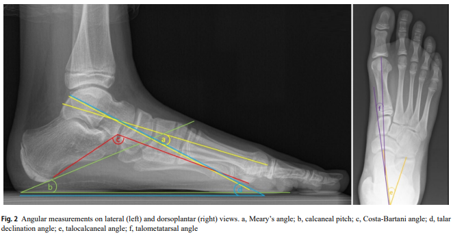
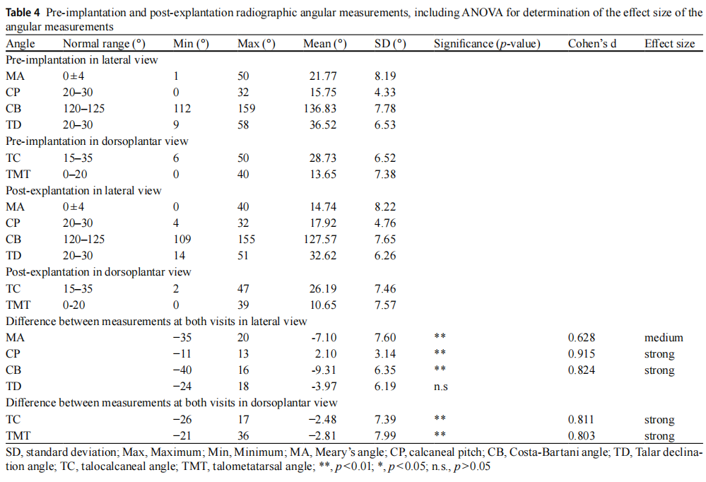
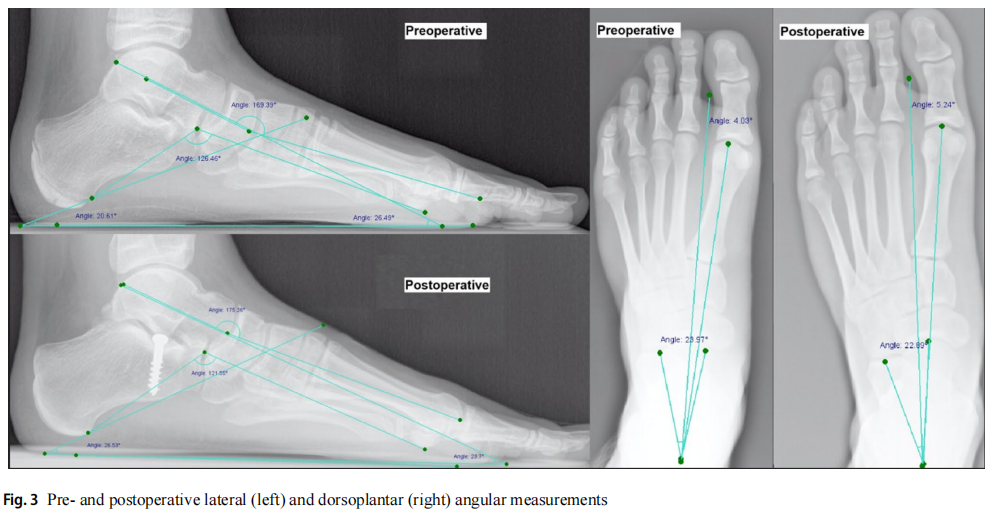
To assess the degree of correction, six different angles (four lateral, and two dorsoplantar) were measured on the preimplantation and pre-explantation radiographs by two of the authors (intra-class correlation coefficient (ICC) = 0.98 using the CentricityTM Universal Viewer (version 6.0, GE Healthcare): Meary's angle (MA) (normal range: 0° ± 4°), calcaneal pitch (CP) (normal range: 20°–30°), Costa-Bartani angle (CB) (normal range: 120°–125°), and talar declination angle (TD) (normal range: 20°–30°) measured on lateral views, and talocalcaneal angle (TC) (normal range: 15°–35°), and talometatarsal angle (TMT) (normal range: 0°–20°) measured on dorsoplantar views (Fig. 2). The raters were blinded to the time-point (pre-implantation vs. pre-explantation) of the radiographs to minimize bias.
Statistical analyses
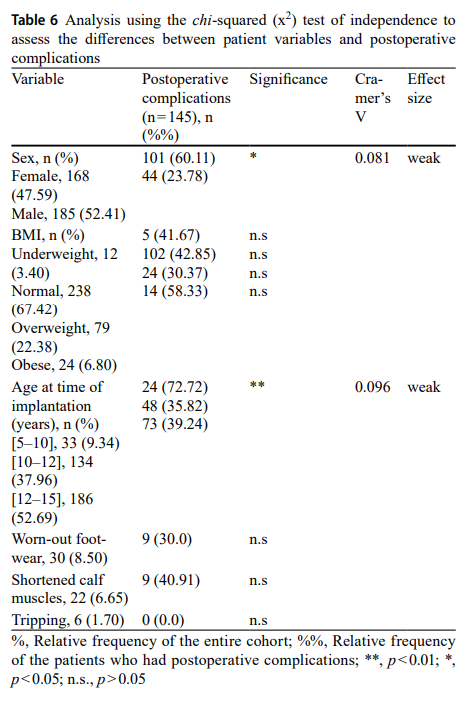
Our institution's electronic medical records system was utilized to collect patient demographics (age, sex, body mass index (BMI)), preoperative factors regarding treatment and pain, data of the SSA, and radiographic images. Quality metrics consisted of postoperative clinical and radiographic outcomes. Other than descriptive statistics, the statistical model consisted of two primary tests: 1) chi-squared (χ²) test of independence to assess the differences between categorical variables and clinical improvement as well as postoperative complication, and 2) Student's t-test (or ANOVA where applicable for multiple groups) to evaluate the change in continuous variables (angular measurements) effect on clinical improvement. Due to the retrospective nature of the study, detailed severity grading and exact duration of each complication were not consistently available for all cases. However, 'resolved' was defined as patient-reported improvement and being pain-free following the specified non-surgical treatments. Results were reported with 95% confidence intervals (CI) to indicate the precision of estimates. In cases where a significant p-value was calculated, Cramer's V (for categorical) and Cohen's d (for continuous parameters) were reported to demonstrate the effect size of the finding: ≤0.1 (V) and ≤0.2 (d) (weak association), 0.1–0.3 (V) and 0.2–0.6 (d) (moderate association), ≥0.5 (V) ≥0.6 (d) (strong association). Given that 175 out of 178 patients underwent bilateral SSA, we acknowledge the potential for within-patient correlation. While the descriptive statistics are presented at the foot level, statistical significance for inferential analyses was interpreted with this potential for non-independence in mind. The significance level was set at p < 0.05 and Bonferroni correction was implemented for adjusting p-values due to the increased risk of a type I error when making multiple statistical analyses. While Bonferroni correction was applied to mitigate the risk of Type I errors from multiple comparisons, we acknowledge that multivariate models could offer a more comprehensive adjustment for confounding variables such as age, sex, BMI, and follow-up time. This will be considered in future prospective studies. All statistical analyses were performed using IBM SPSS software version 23 (IBM Corporation, Armonk, New York, United States). All variables included in the analyses had complete data, with no missing values.
Results
Patient demographics and primary outcomes
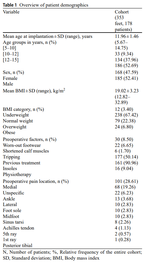
The data on 178 patients (353 feet) (168 (47.59%) females, and 185 (52.41%) males) who underwent SSA between December 2007 and July 2018 were meticulously analyzed. The mean follow-up time was 51.47 ± 20.21 months (range 4–135 months). 175 patients had bilateral SSA, 141 of which were operated on in the same setting, and 34 were operated on at another date. Preoperatively, 30 (8.50%) feet had worn-out footwear. Shortened calf muscles were in 22 (6.65%) feet, and tripping was observed in 6 (1.70%) feet. Pain was mainly localized medially (28.61%). 177 (51.14%) feet had specific previous non-surgical treatment; of which, 161 (90.96%) were treated with foot insoles, and 16 (9.04%) with physiotherapy.
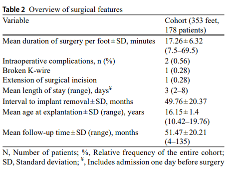
At the time of implantation, the mean age was 11.96 ± 1.46 (5.67–14.75) years, and the mean BMI was 19.02 ± 3.23 (12.82–32.89) kg/m². The majority of the patients (67.42%) had normal BMI values, 22.38% were overweight, 6.80% were obese, and 3.40% were underweight. The mean length of the implantation surgery per foot was 17.26 ± 6.32 (7.5–69.5) minutes. 99.44% of the implantation surgeries did not have any complications. One foot had an intraoperative complication of a broken K-wire, which was left in situ, and another foot required extension of the surgical incision. Following the implantation surgery, patients were hospitalized for a period between two and eight days, with a mean of three days. It is essential to highlight that several patients were admitted one day before surgery. The implant was removed 49.76 ± 20.37 months following SSA, without any reported complications. At the time of the removal, patients' age was 16.15 ± 1.4 (10.42–19.76) years.
Clinical improvement
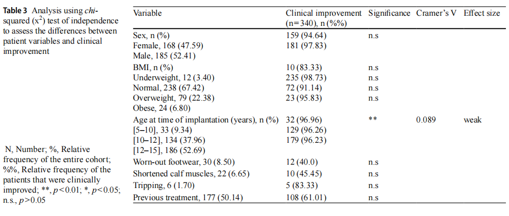
At the first postoperative outpatient visit, 340 (96.31%) feet were found to have clinician-reported improvement as reduction in pain and discomfort was reported, foot pronation and hindfoot valgus were corrected, and the medial arch was improved. Statistical analysis demonstrated that the younger age group [5–10] years exhibited a weak, yet significant clinical improvement when compared to the older groups (96.96% vs. 96.26% and 96.23%, p < 0.01, V = 0.089). Patient's sex, BMI, worn-out footwear, shortened calf muscles, tripping, and previous treatment status demonstrated no significant effect on the clinical improvement.
Radiographic correction
Meary's angle (MA)
Prior to SSA, mean MA was 21.77° ± 8.19° (1°–50°). After explantation, values reduced to 14.74° ± 8.22° (0°–40°), difference -7.1° ± 7.6°.
Calcaneal pitch (CP)
Increased by 2.1° ± 3.14°, from 15.75° ± 4.33° (0°–32°) to 17.92° ± 4.76° (4°–32°).
Costa-Bartani angle (CB)
Decreased by 9.31° ± 6.35°, from 136.83° ± 7.78° (112°–159°) to 127.57° ± 7.65° (109°–155°).
Talar declination angle (TD)
Decreased by 3.97° ± 6.19°, from 36.52° ± 6.53° (9°–58°) to 32.62° ± 6.26° (14°–51°).
Talocalcaneal angle (TC)
Reduced by 2.48° ± 7.39°, from 28.73° ± 6.52° (6°–50°) to 26.19° ± 7.46° (2°–47°).
Talometatarsal angle (TMT)
Decreased by 2.81° ± 7.99°, from 13.65° ± 7.38° (0°–40°) to 10.65° ± 7.57° (0°–39°).
Following treatment, five of the six (83.33%) measured angles demonstrated values approaching the normal range. All angular measurements except TD demonstrated medium-to-strong (d = 0.628–0.915) effect sizes. CP exhibited the strongest effect size (d = 0.915, 95% CI 0.850–0.980).
Postoperative complications
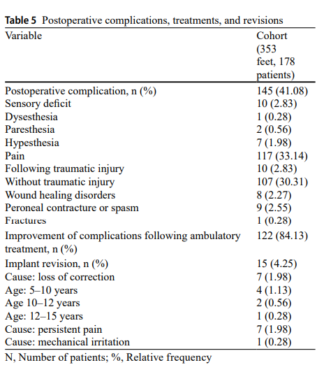
At the first postoperative ambulatory follow-up, 41.08% of the cohort demonstrated complications. The vast majority were due to postoperative pain (33.14%). 2.83% had sensory deficits, 2.55% peroneal contracture or spasm, 2.27% wound healing disorders, and 0.28% fractures. Following treatment, 84.13% of patients reported improvement and being pain-free. 4.25% required implant revision due to loss of correction, persistent pain, or mechanical irritation.
Discussion
The Ilizarov method is recommended by a number of authors for treating nonunion of the tibia, as it is highly effective in achieving bone union, treatment of a possible infection, correcting limb length discrepancy and axial misalignment, and eliminating joint contractures1,2,3,4,5,7,8,9,10,11,12,13,14,15,17,18,19.
There are various recommended treatment strategies of nonunions of the tibia employing the Ilizarov method2,3,4,5,7,8,9,10,11,12,13,14,15,16,17,19,20,21,22. Good treatment outcomes have been demonstrated with the use of various treatment strategies2,3,4,5,6,7,8,11,12,13,14,15,16,18,19,20. These include: fixation alone9,12,15,17,18; fixation and compression7,20; fixation, segmental resection, and bone transport2,4,6,12,14,15,19,20,21; and fixation, resection, and compression with bone transport5,7,8,10,11,13,14,20. Eralp observed good treatment outcomes in infected nonunions of the tibia treated by means of either combined fixation and compression or combined fixation, resection, and compression with bone transport7. However, various surgical techniques and treatment strategies may affect the outcomes in ways that are not known at this time. McNally et al. assessed the effect of four different treatment strategies and techniques used in infected pseudarthrosis of the tibia on treatment outcomes in 79 patients20. These strategies and techniques were: monofocal distraction, monofocal compression, bifocal compression/distraction, and bone transport20. Post-treatment infection recurrence was observed in three patients from the monofocal compression subgroup. Primary bone union rates were the lowest (73.7%) in the monofocal compression subgroup and the highest in the bifocal compression/distraction (93.8%) and monofocal distraction (96.2%) subgroups. The authors concluded by advising against the use of monofocal compression in the treatment of pseudarthrosis of the tibia20.
Our study demonstrated bone union in 100% of patients, which is an outcome comparable to, or even slightly better than, those reported in literature (73.7–100%) Table71,2,3,4,5,6,7,8,9,10,11,12,13,15,16,17,18,19,20,21,22. The treatment strategy and surgical technique showed noeffecton the proportion of patients who achieved bone union in the individual subgroups.
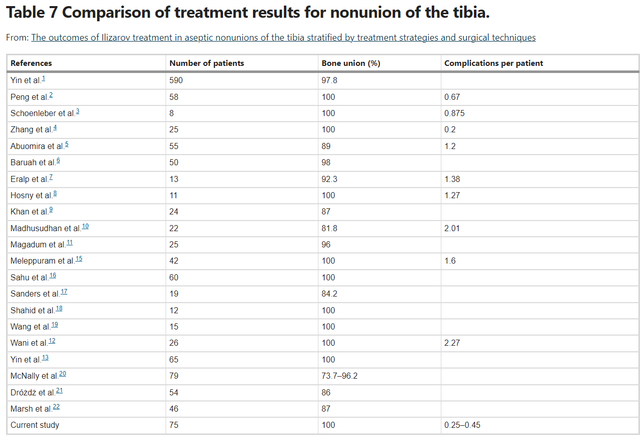
There have been no studies assessing the number of complications depending on the employed treatment strategy and surgical technique. Our study population, depending on the subgroup, developed anywhere from 0.25 to 0.47 complications per patient, which is a slightly better result than those reported in literature, which range from 0.67 to 2.27, Table72,3,4,19. The employed treatment strategies and surgical techniques were observed to have no effect on the mean number of complications per patient.
There are no available reports from studies assessing the duration of treatment with an external fixator stratified by different treatment strategies and surgical techniques. Overall, the mean duration of treatment ranges from 5.8 months to 13.5 months2,3,4,5,6,8,9,16,19. These figures are similar to ours. We observed no effect of the evaluated treatment strategies or surgical techniques on Ilizarov treatment duration.
ASAMI bone scores reported by Abuomira were 51% excellent, 33% good, 9% fair, and 7% poor5. Khan observed 25% excellent, 58.3% good, 4.2% fair, and 12.5% poor ASAMI bone scores9. Meleppuram achieved 60% excellent, 15% good, and 25% fair ASAMI bone scores15. None of the authors cited here assessed the ASAMI bone scores stratified by the employed treatment strategy and surgical technique.
The treatment strategies and surgical techniques employed in our evaluated patient population yielded no significant differences in the resulting ASAMI bone scores.
The ASAMI functional scores reported by Abuomira were 45% excellent, 38% good, 9% fair, and 7% poor5. Khan observed 33.3% excellent, 50% good, 8.35% fair, and 8.35% poor ASAMI functional scores9. Meleppuram reported 55% excellent, 30% good, 5% fair, and 10% poor ASAMI functional scores15. The relevant literature contains no studies assessing ASAMI functional scores stratified by the employed surgical technique and treatment strategy.
In our study, treatment strategy was observed to have no effect on the ASAMI functional score. However, when it comes to surgical techniques, the patients who underwent closed fixation achieved significantly higher ASAMI functional scores than the open-fixation patients. This may be a result of better soft-tissue and surgical-wound healing.
We are aware of the fact that treatment of infected and aseptic pseudarthroses may produce different outcomes. Unfortunately, there is a scarcity of papers addressing aseptic pseudarthrosis treatment in the available relevant literature. Our study is one of the first ones to analyze the available techniques and strategies employed in treating pseudarthroses with an Ilizarov fixator.
The available literature reports inform us that the mean length of hospital stay for treating patients with nonunions of the tibia with an external fixator ranges from 5 to 105 days4,12,16. These statistics are slightly worse than those achieved in our study. We observed no effect of the employed surgical technique or treatment strategy on the length of hospital stay.
The most common complication observed in our study population during treatment with an Ilizarov fixator was Kirschner wire pin tract infection. Such infections typically respond well to topical antiseptics and oral antibiotic therapy in an outpatient setting. Deep infections involving soft tissues and bone require hospitalization, surgical debridement, and Kirschner wire replacement, which significantly lengthens the healing process (median: 189.0 days vs. 248.5 days).
Bone transport is a more complex procedure than that of employing compression/distraction. There may be problems with achieving good contact of bone ends and ensuring bone union at the docking site; moreover, more complications may develop, and the duration of treatment may be longer5. Open fixation procedures in patients with nonunions are more complex than closed fixation. Continued compression is more bothersome for nonunions patients than neutral fixation without compression.
We observed better ASAMI functional score outcomes in the patients who underwent closed fixation than in the open fixation group.
The different surgical techniques had no effect on the number of complications, rates of bone union, length of hospital stay, duration of Ilizarov treatment, or ASAMI bone scores.
The different treatment strategies had no effect on the number of complications, rates of bone union, rates of bone union, length of hospital stay, duration of Ilizarov treatment, ASAMI bone scores, or ASAMI functional scores.
Multicenter, randomized studies are needed in order to compose the guidelines for the treatment of aseptic pseudarthroses of the tibia. Nonetheless, our study can be considered an attempt to assess various techniques and strategies in the treatment of tibial nonunion and present our team’s experiences.
For managing nonunions of the tibia we recommend the technique of closed fixation without continued compression.
Nonetheless, the use of the Ilizarov method in the treatment of nonunions of the tibia yields good outcomes irrespective of the employed surgical technique or treatment strategy.
Limitations
Retrospective design, potential non-independence of bilateral feet, lack of control group, absence of validated patient-reported outcome measures, limited dynamic imaging, weak effect sizes for some significant findings, and institution-specific postoperative protocol.
Conclusions
SSA is a minimally invasive technique associated with significant clinical improvement and radiographic correction in pediatric FF. Younger age at surgery correlated with better outcomes. Complication rate was high but mostly minor and manageable. Careful patient selection is essential.
References
(Full reference list available in original document)
Declarations
Conflict of interest: The authors declare no conflict of interest.
Open Access: This article is licensed under a Creative Commons Attribution 4.0 International License.