EN
EN


Characteristics and Surgical Techniques of the "Double‑Intra" Type of Irreducible Intertrochanteric Fractures
Intertrochanteric fractures are one of the three major osteoporotic fractures, with high incidence, high disability rates, and high rates of internal fixation failure. Closed reduction and intramedullary nailing is currently the first‑line treatment for intertrochanteric fractures. Most of these fractures can achieve satisfactory reduction with traction combined with internal/external rotation. However, some special fracture types are difficult to reduce closed and require open reduction, and are often termed "irreducible" intertrochanteric fractures.
In 2025, the latest issue of the Journal of Orthopaedic Surgery and Research (JOSR) published a study by the orthopaedic team from Yangpu Hospital, Tongji University, Shanghai, on a specific type of intertrochanteric fracture characterized by difficulty in reduction and a tendency for redisplacement during and after surgery.
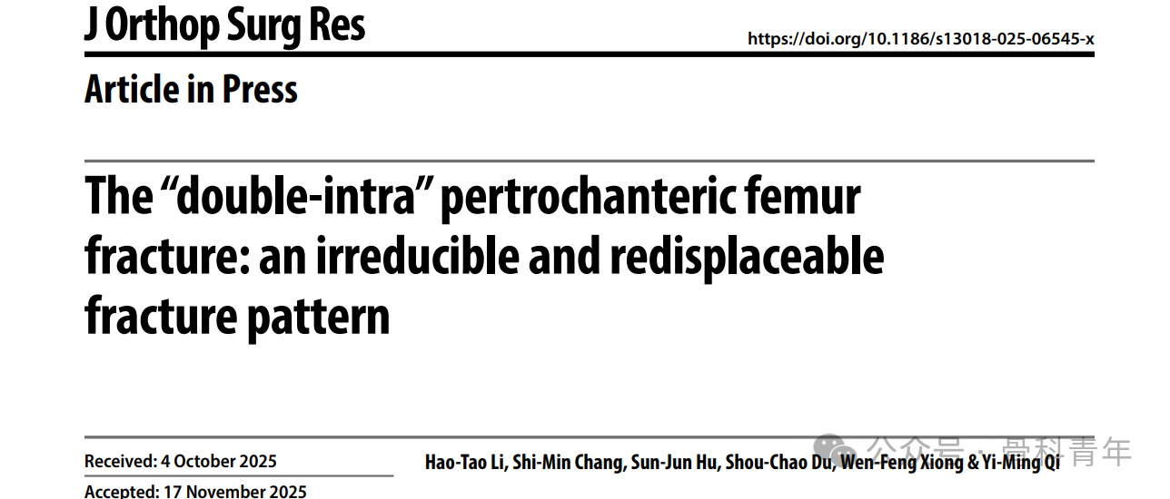
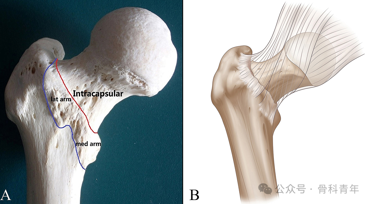
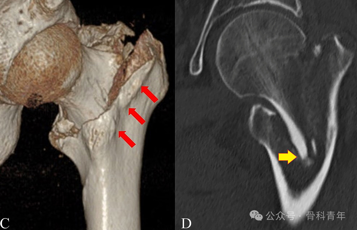
The team named this specific fracture type the "double‑intra" intertrochanteric fracture. The two "intra" components refer to:
- Intracapsular – the anterior fracture line of the intertrochanteric fracture lies within the joint capsule;
- Intramedullary – the head‑neck fragment is impacted into the medullary cavity.
Study Results
A total of 28 patients were included in this study, with the following results:
1. Intraoperative and postoperative changes in reduction quality
Using the reduction quality criteria proposed by Professor Zhang Shimin (Chang criteria), all 28 patients achieved an "excellent" reduction (score 4) before nail insertion: 16 via manual manipulation and 12 with bone‑hook‑assisted lever reduction. After intramedullary nailing, reduction quality dropped to "acceptable" (score 3 or 2) in 10 patients and to "poor" in 3 patients.
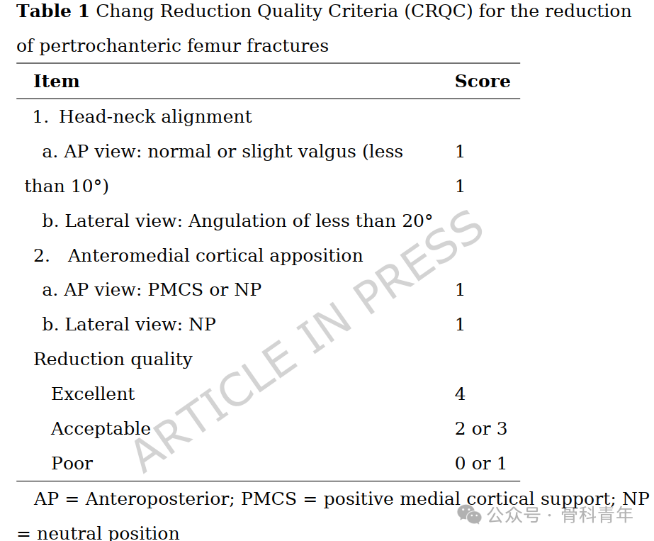
The rate of reduction loss due to nail insertion was 46.4% (13/28). Postoperative 3D CT showed that only 8 patients (28.6%, 8/28) achieved anteromedial cortical support, while 20 patients (71.4%, 20/28) completely lost anteromedial support. Compared with immediate postoperative fluoroscopic images, 7 of 15 patients (46.7%, 7/15) showed further loss of anteromedial cortical support on follow‑up.
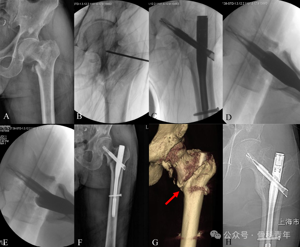
In the patient shown above, anteromedial cortical support was maintained after temporary K‑wire fixation followed by nail insertion, and was still present at 1.5‑year follow‑up.
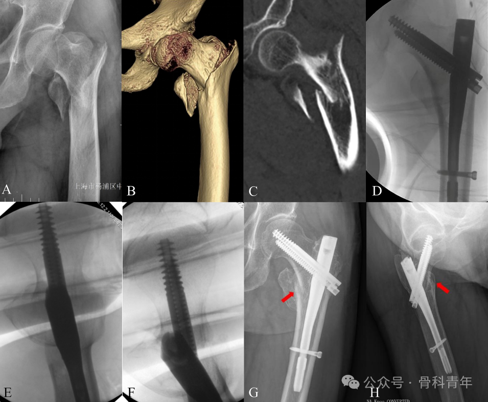
The patient above achieved neutral support immediately postoperatively, but lost anteromedial cortical support at 18‑month follow‑up.
2. Effect of temporary K‑wire fixation
A total of 9 patients received temporary K‑wire fixation intraoperatively. In 2 of these, the K‑wire had to be removed during surgery because it obstructed nail insertion. Overall, among these 9 patients, 1 experienced reduction loss after nail placement, while the remaining 8 maintained reduction.
In contrast, among the 19 patients without temporary K‑wire fixation, 12 lost cortical support after nail insertion and 7 maintained it. The intraoperative reduction loss rate in the non‑K‑wire group (63.2%, 12/19) was significantly higher than that in the K‑wire group (11.1%, 1/9).
Discussion
1. Relationship between fracture characteristics and difficult reduction
Anatomically, the trochanteric region is the junction between the femoral neck and the metaphysis, with several specific features. First, the femoral neck is relatively narrow and composed mainly of dense cortical bone, whereas the distal trochanteric cross‑section is wider and consists mainly of cancellous bone. Consequently, the head‑neck fragment tends to slide into the wider medullary cavity of the femoral shaft and become locked by bone tissue. Second, the intertrochanteric line is a prominent ridge at the junction of the neck and the anterior surface of the shaft. The medial arm of the iliofemoral ligament passes over the anterior surface of the femoral head and attaches to the lower part of the intertrochanteric line. Classical textbooks and extensive literature describe intertrochanteric fractures as extracapsular. However, the anterior fracture line of intertrochanteric fractures is most commonly extracapsular or transcapsular, while a small proportion are completely intracapsular. In intracapsular fractures, the anterior capsule may act as a soft‑tissue barrier, limiting anterior displacement of the fragment and thus impeding anterior reduction.
2. Relationship between fracture characteristics and redisplacement tendency
We observed that this fracture subtype is highly prone to loss of cortical support both during and after surgery, even after initial satisfactory reduction. Based on the specific anatomical features of the "double‑intra" fracture, we propose the following biomechanical explanations:
1) The initial intramedullary impaction of the head‑neck fragment compresses the cancellous trabeculae, leaving a large void in the metaphyseal medullary cavity after reduction. This void provides no mechanical resistance to the head fragment, leading to subsequent subsidence and eventual re‑impaction into the medullary cavity. This is particularly problematic in elderly patients, where severe osteoporosis significantly reduces the fixation capacity of cancellous bone and implants, accelerating secondary displacement.
2) Gravity in the supine position, combined with continuous tension from the posterior capsule, may cause posterior displacement of the anterior cortex of the head‑neck fragment.
3) Due to the limited resolution of intraoperative fluoroscopy, a 2‑mm cortical step may not be appreciated during surgery, and mild malalignment may be misinterpreted as neutral alignment.
4) Postoperative muscle contraction and early mobilization may contribute to secondary displacement. This dynamic instability can ultimately lead to poor cortical support, gradual collapse, and implant failure.
3. Compensatory techniques to enhance stability
Achieving satisfactory reduction—especially obtaining positive anteromedial cortical support—is crucial for fracture stability. Therefore, we recommend using a bone hook to elevate the head‑neck fragment, deliberately creating "double‑positive" support of both the medial and anterior cortices. In particular, positive anterior cortical support acts as a mechanical buffer to accommodate potential postoperative reduction loss. Importantly, this target position is easily visualized intraoperatively: a 30° oblique lateral view should show overlap of the distal anteromedial cortex with the proximal fragment tip. This reduction technique does not require direct exposure of the fracture site; it can be performed through a small incision, with the bone hook guided by finger palpation without direct visualization. This approach avoids extensive soft‑tissue dissection, preserves the surrounding vascular structures, and minimizes disruption of the fracture haematoma and blood supply.
However, due to the "void" created by cancellous bone impaction, the head‑neck fragment tends to slide into the femoral medullary cavity. Our findings show that the reduction loss rate was significantly higher in the group without temporary K‑wire fixation compared with the K‑wire group. During surgery, cortical support is very fragile and difficult to maintain. Therefore, it is essential to use 1‑2 K‑wires for temporary fracture fixation before nail insertion to prevent intraoperative reduction loss.