EN
EN


Open Reduction and Internal Fixation of Hoffa Fracture with Plate and Screws
1. Principles
General Considerations
Hoffa fractures involve the critical weight-bearing surface of the knee joint and require anatomical reduction and absolute stability of fixation. In general, a buttress plate with lag screws is recommended. However, if the fracture fragment is small, lag screws alone may be the only feasible fixation method. Indirect anterior lag screw technique is not recommended, as the long screw threads fail to provide adequate stability and effective compression of the fracture fragments. The same principles apply if both condyles are fractured.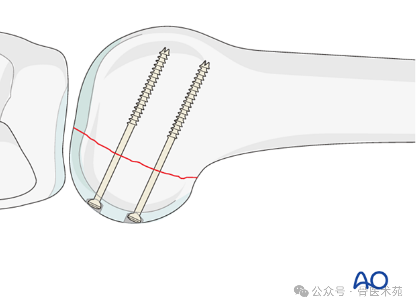
Screw Types
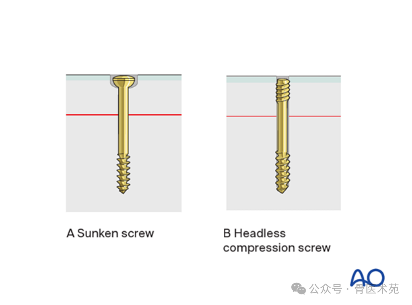
All implants must not protrude above the articular surface. This can be achieved using countersunk lag screws (A) or headless compression screws (B). To prevent fragment rotation, at least two screws should be used. In this procedure, 3.5 mm cannulated headless compression screws or standard 3.5 mm lag screws may be selected. However, alternative screw sizes may be used depending on fragment dimensions.
2. Patient Positioning and Surgical Approach
Patient Positioning
The patient is placed in a supine position with the knee flexed 20°–30°.
Approach
- For lateral Hoffa fractures, the Swashbuckler approach or Gerdy's tubercle osteotomy approach may be used.
- For medial Hoffa fractures, the internervous medial approach is employed.
3. Reduction
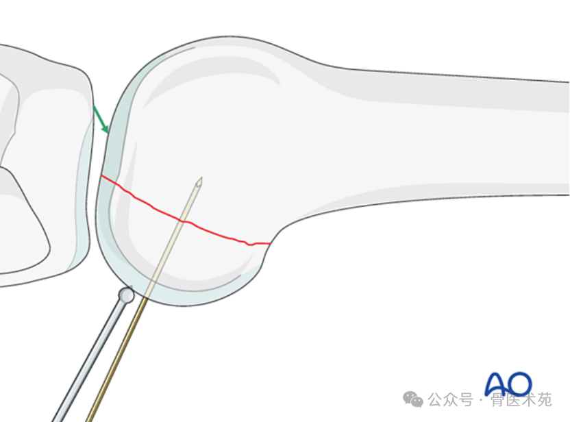
The fracture is reduced using a small ball-tipped push‑rod and temporarily fixed with K‑wires. Ensure that K‑wire placement does not interfere with the planned plate position or screw trajectories.
4. Fixation
Principles
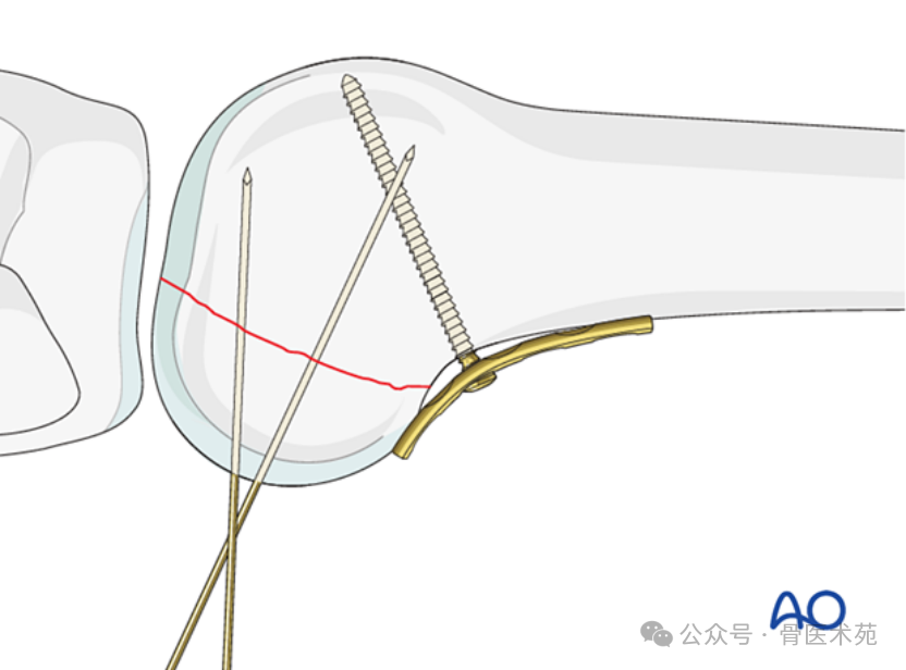
To enhance stability and avoid axial loading across the fracture site (particularly in osteoporotic bone), a buttress plate is used to prevent proximal migration of the fragment. The plate is placed according to the location of the unstable fragment. Occasionally, the fragment may be located laterally. Various plate types are available; for example, a low‑profile 3.5 mm narrow plate is used here as an illustration.
Plate Application
The plate is applied to the posterior aspect of the distal femur. It should be placed as distal as possible without impinging on the articular surface. To achieve close apposition of the low‑profile plate to the femur, a standard cortical screw is inserted proximal to the fracture line in neutral mode.
Screw Insertion in Neutral Mode
Proximal to the first screw, one or more bicortical screws are placed to secure the plate. If screws can be placed in non‑articular areas, additional screws may be inserted in the distal part of the plate. All screws are inserted in neutral mode.
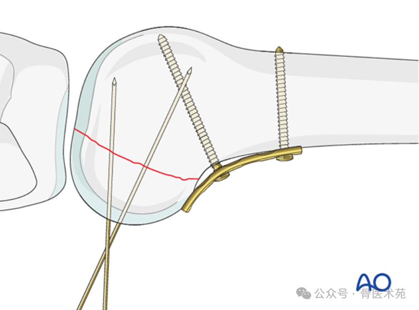
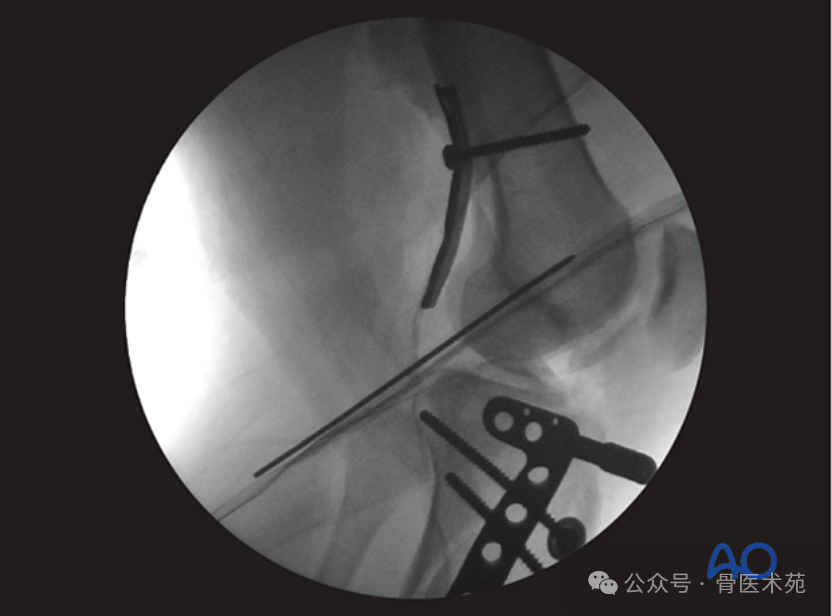
Intraoperative image showing reduction of a posterior Hoffa fracture, temporary K‑wire fixation, and posterior buttress plate reinforcement.
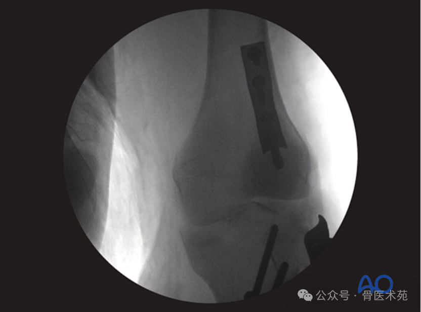
Postoperative radiograph showing the posterior buttress plate properly positioned.
Guide Wire Placement
Two guide wires are inserted perpendicular to the fracture plane, ensuring they do not penetrate the far cortex.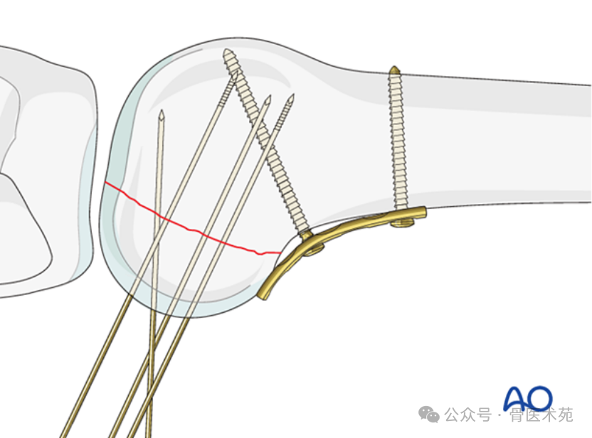
Checking Guide Wire Position
Guide wire position is verified under image intensification in lateral and oblique views.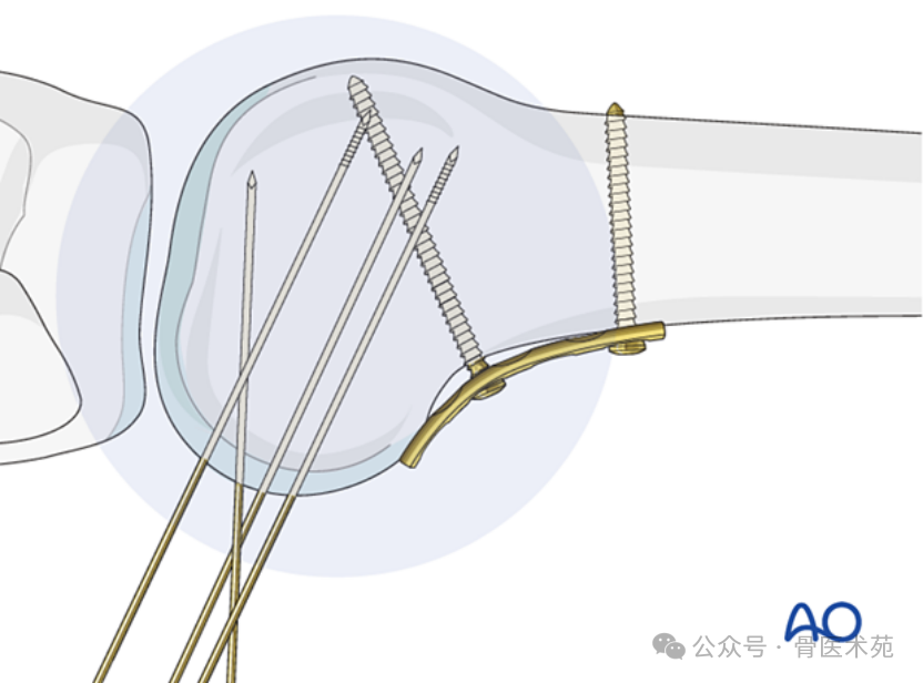
Headless Compression Screw Insertion
Headless compression screws are inserted using a cannulated screwdriver, and screw length is checked under image‑intensified lateral view.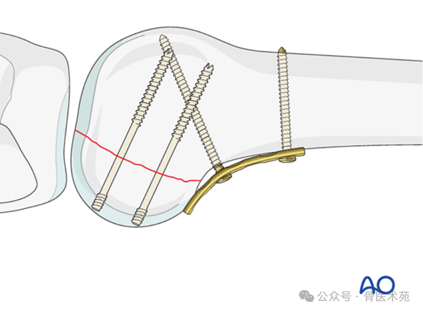
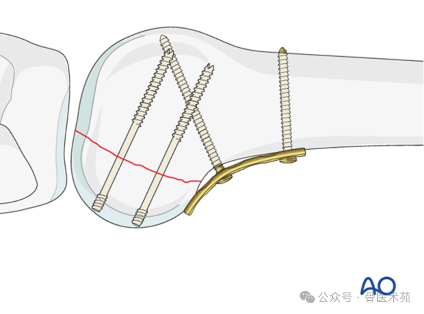
Cancellous Lag Screw Insertion (Alternative: Standard Screws)
Under image intensification, lag screws are inserted according to standard cancellous lag screw technique. Countersinking is performed to avoid screw head prominence.
5. Case Example
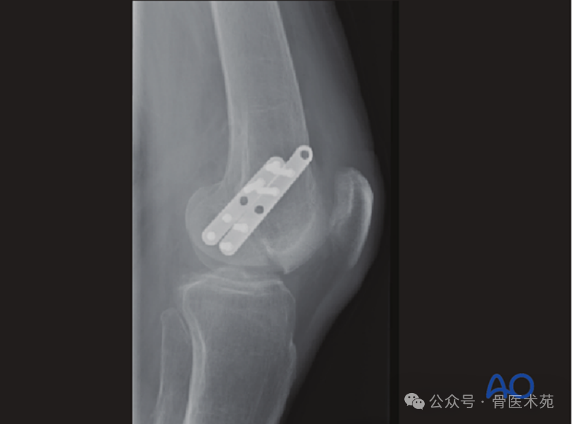
As illustrated, if the plate is placed laterally or medially rather than posteriorly, fixation failure is likely. This image shows articular surface incongruity, which would require revision surgery.
6. Postoperative Rehabilitation
After distal femoral fractures, the main obstacles to full knee function recovery include fibrosis and adhesions of the soft tissues surrounding the metaphyseal fracture zone, capsular scar formation, intra‑articular adhesions, and muscle weakness. Early range‑of‑motion exercises help restore joint motion in the early postoperative period. With stable fracture fixation, the surgeon and physical therapist will design a progressive rehabilitation program tailored to each patient. The recommendations here are for reference and are not mandatory.
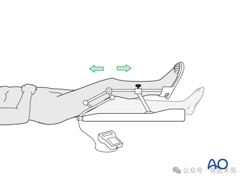
Functional Therapy
Unless other injuries or complications exist, knee mobilization can begin immediately after surgery. Active and passive movements of the knee and hip can be initiated on the first postoperative day. Therapy should focus on progressive quadriceps strengthening and straight‑leg raises. Unloaded stationary cycling, along with firm but passive range‑of‑motion exercises, helps the patient achieve optimal joint mobility.
Weight‑bearing
Touch‑down weight‑bearing (10–15 kg) with crutches or a walker can commence immediately after surgery. This is continued for 6–10 weeks, primarily to protect the injured joint rather than the diaphyseal region. From 6–10 weeks postoperatively, touch‑down weight‑bearing is progressively transitioned to full weight‑bearing over 2–3 weeks. Ideally, the patient should achieve full weight‑bearing without assistive devices by 12 weeks.
Follow‑up
Wound healing is assessed at 2–3 weeks. Subsequent follow‑ups are typically scheduled at 6 weeks, 12 weeks, 6 months, and 12 months. Serial radiographs allow the surgeon to evaluate fracture healing progress.
Implant Removal
Implant removal is not mandatory, but if implant‑related symptoms develop after fracture union, removal should be discussed with the patient.
Thromboembolic Prophylaxis
Thromboprophylaxis should follow local treatment guidelines.