EN
EN


ক্যালকেনিয়াস ফ্র্যাকচারের জন্য সর্বনিম্ন আক্রমণাত্মক কে-ওয়্যার এবং স্ক্রু ফিক্সেশন
১. রোগ নির্ণয়
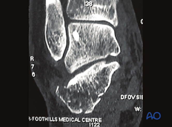
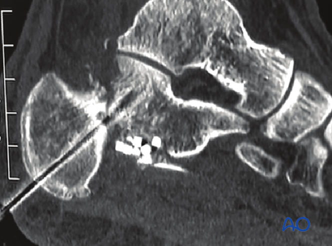
উদাহরণ ছবি: সিটি স্ক্যানে পোস্টেরিয়র ফ্যাসেটের একটি সরে যাওয়া ফ্র্যাকচার দেখা যায়, যা একটি একক ফ্র্যাগমেন্ট হিসেবে বিবেচিত হয় এবং সাবকন্ড্রাল ল্যাগ স্ক্রু দিয়ে ন্যূনতম ইনভেসিভ উত্থাপন ও ফিক্সেশনের জন্য উপযুক্ত।
২. সার্জিক্যাল অ্যাপ্রোচ
সাইনাস টারসি অ্যাপ্রোচ অথবা একটি ন্যূনতম ইনভেসিভ ল্যাটারাল অ্যাপ্রোচ ব্যবহার করা যেতে পারে।
৩. রিডাকশন
অপারেশনের পূর্বে প্রস্তুতি
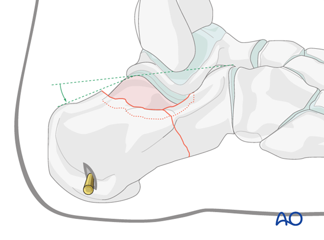
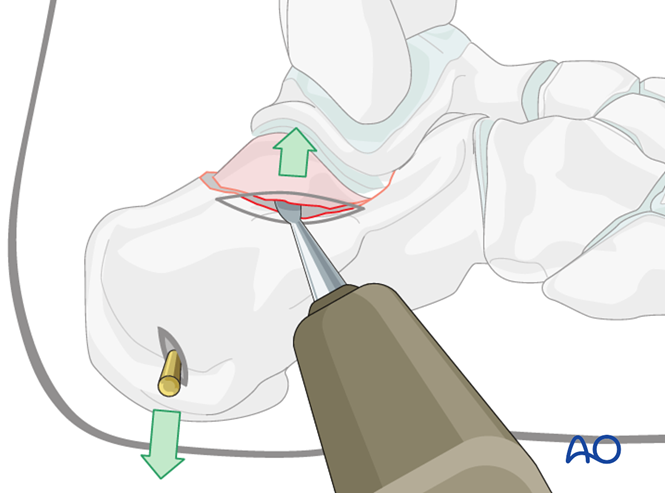
রোগীকে ল্যাটারাল ডেকুবিটাস অবস্থানে স্থাপন করা হয়। ইমেজ ইনটেনসিফায়ার নিয়ন্ত্রণের অধীনে, ক্যালকেনিয়াসের পোস্টেরিয়র টিউবারোসিটিতে একটি স্টেইনম্যান পিন সন্নিবেশিত করা হয় যাতে ক্যালকেনিয়াল দৈর্ঘ্য পুনরুদ্ধারের জন্য জয়স্টিক হিসেবে কাজ করে।
অবসাদিত ফ্র্যাগমেন্টের উত্থাপন
ফ্লোরোস্কোপিক নির্দেশনায়, ফ্র্যাকচারের কেন্দ্রের উপর একটি ছোট চিড় (যেমন: সাইনাস টারসি অ্যাপ্রোচ) করা হয়। একটি পেরিওস্টিয়াল এলিভেটর দিয়ে অবসাদিত ফ্র্যাগমেন্টটি তুলে আর্টিকুলার কনগ্রুয়িটি পুনরুদ্ধার করা হয়।
হ্রাস বজায় রাখা
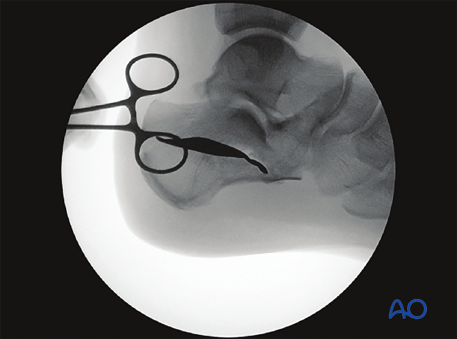
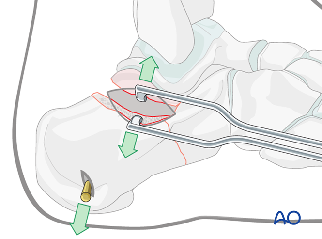
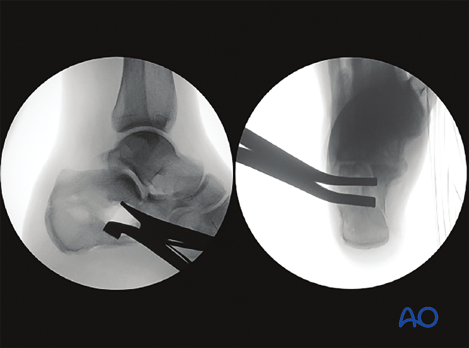
একটি ডিস্ট্রাক্টর ব্যবহার করা হয় রিডাকশন বজায় রাখতে সহায়তা করার জন্য, এবং ফ্লুওরোস্কোপি করা হয় উপযুক্ত রিডাকশন নিশ্চিত করার জন্য।
উদাহরণ ছবি: ফ্লুওরোস্কোপিক দৃশ্য যা ৩ সেমি ইনসিশনের মাধ্যমে ডিস্ট্রাক্টর ব্যবহার করে পোস্টেরিয়র ফ্যাসেটের উত্থান দেখায়।
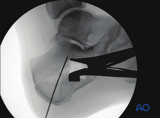
তারপর পার্কিউটেনিয়াস কে-ওয়্যার বা স্ক্রু স্থাপন করা হয় রিডাকশন ধরে রাখার জন্য; ফ্র্যাগমেন্টগুলিকে স্থিতিশীল করতে এবং নিরাময় প্রবর্তন করতে ডিফেক্টে বোন সাবস্টিটিউট প্যাক করা যেতে পারে।
৪. ফিক্সেশন
কে-ওয়্যার ফিক্সেশন
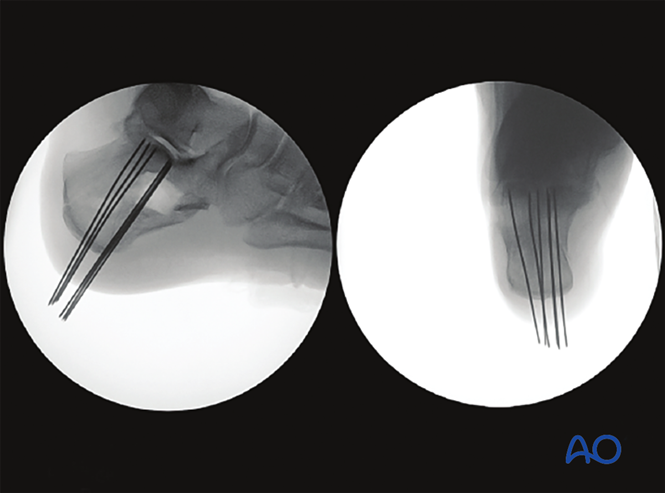
অপারেশনের পরের এক্স-রে বোহলার’স অ্যাঙ্গেলের পুনরুদ্ধার দেখায়, যেখানে কে-ওয়্যারগুলি পোস্টেরিয়র টিউবারোসিটি থেকে পোস্টেরিয়র ফ্যাসেটে প্রবেশ করানো হয়েছে এবং ডিফেক্টটি বোন সাবস্টিটিউট দিয়ে পূর্ণ করা হয়েছে (মাত্র ২ সেমি ইনসিশনের মাধ্যমে)।
ছবিগুলি দুটি প্রোজেকশনে কম হওয়া পোস্টেরিয়র ফ্যাসেটকে দেখায়, যা কে-ওয়্যার দিয়ে ফিক্স করা হয়েছে।
স্ক্রু বা প্লেট ফিক্সেশন
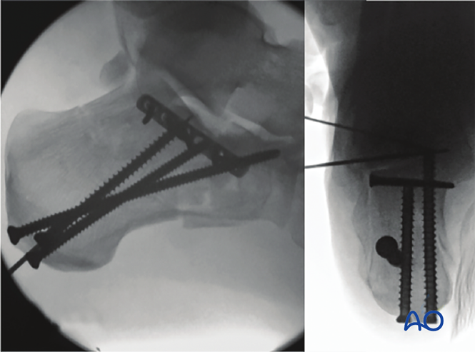
কে-ওয়্যার ছাড়াও, পার্কিউটেনিয়াস স্ক্রু বা প্লেট ফিক্সেশনের জন্য ব্যবহার করা যেতে পারে।
৫. অপারেশনের পরের ব্যবস্থাপনা
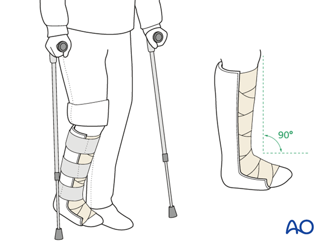
শুরুতে, আংকেলকে নিউট্রাল অবস্থানে রাখতে একটি ঘন পোস্টেরিয়র স্প্লিন্ট প্রয়োগ করা হয়, এবং আক্রান্ত অঙ্গটিকে মাঝে মাঝে উত্থিত করা হয়।
গোড়ালির গতিসীমা ব্যায়াম অপারেশনের ২–৫ দিন পর থেকে শুরু করা হয়। পিন-ট্র্যাক্ট সংক্রমণ প্রতিরোধের জন্য নিয়মিত পরিষ্কার করা হয়।
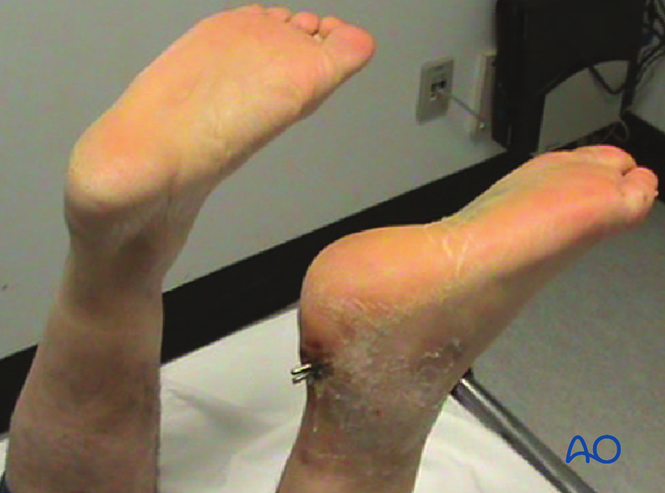
কে-ওয়্যার অপসারণ: অপারেশনের ৬ সপ্তাহের আগে করা হবে না (উদাহরণ ছবিতে ৬ সপ্তাহ পর কে-ওয়্যারের অবস্থা দেখানো হয়েছে)।
ওজন বহন: ৬ সপ্তাহ পর পিন অপসারণের পর থেকে শুরু করা হবে।
চিত্র অনুসরণ: অপারেশনের ৬, ১২ ও ২৬ সপ্তাহ পর পার্শ্বীয় ও অক্ষীয় এক্স-রে করা হয়।