EN
EN

Minimálisan invazív K‑drót- és csavarrögzítés sarokcsont-törések esetén
1. Diagnózis
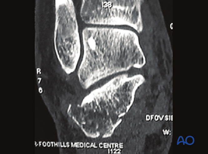
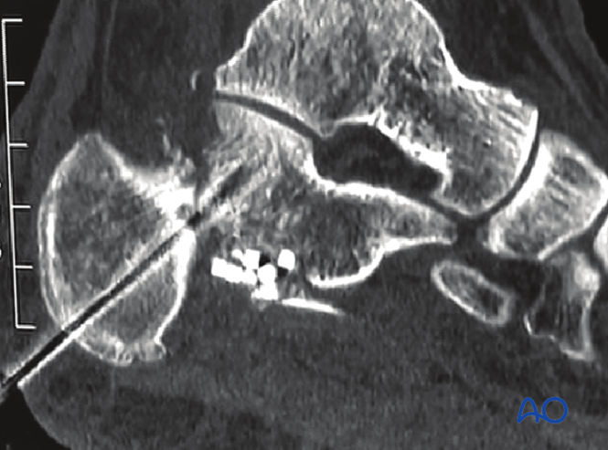
Példakép: A CT a hátsó ízfelszín elmozdult törését mutatja egyetlen töredékként, amely minimálisan invazív emelésre és subchondralis lag-csavarral történő rögzítésre alkalmas.
2. Sebészi megközelítés
A sinus tarsi megközelítés vagy egy minimálisan invazív oldali megközelítés is alkalmazható.
3. Redukció
Műtét előtti felkészülés
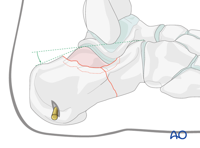
A beteget oldalfekvésbe helyezzük. Képintenzitás-fokozó irányítás alatt egy Steinmann-tűt vezetünk be a sarokcsont hátsó tuberositásába, hogy „joystick”-ként szolgáljon a sarokcsont hosszának visszaállításához.
A süllyedt töredék emelése
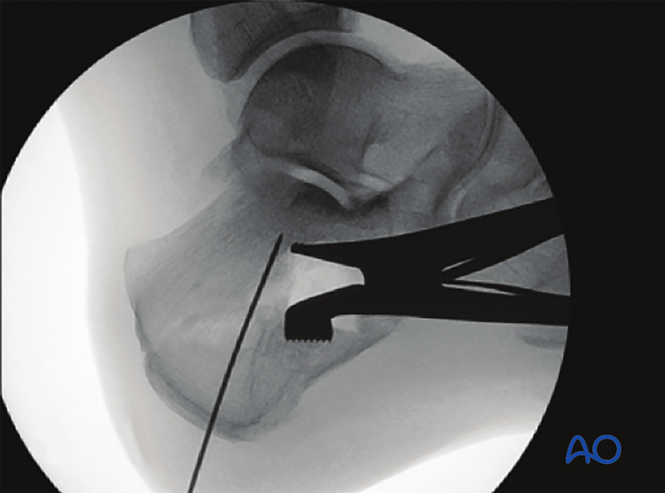
Fluoroszkópiai irányítás mellett egy kis metszést (pl. sinus tarsi megközelítés) hoznak létre a törés középpontja fölött. Egy periostealis emelőt helyeznek be, hogy felemeljék a lenyomódott töredéket, és helyreállítsák az ízületi felületek illeszkedését.
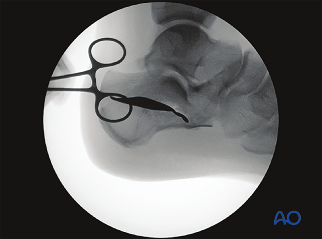
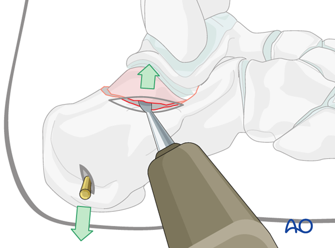
A redukció megtartása
A redukció megtartásához distraktort használnak, és fluoroszkópiát végeznek a megfelelő redukció megerősítésére.
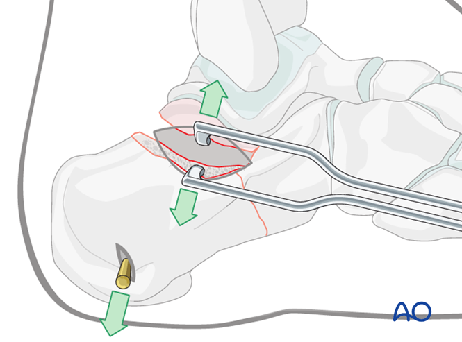
Példakép: Fluoroszkópiai kép, amely a distraktor segítségével történő hátsó ízületi felület felemelését mutatja egy 3 cm-es metszésen keresztül.
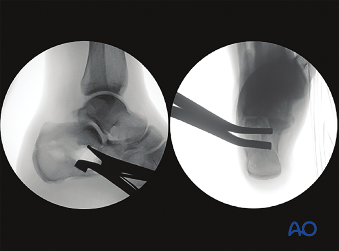
Ezután percután K-drótokat vagy csavarokat helyeznek el a redukció rögzítésére; a hiányosságba csontpótló anyagot is be lehet tömni a töredékek stabilizálására és a gyógyulás elősegítésére.
4. Rögzítés
K-drót rögzítés
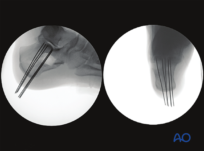
Műtét utáni röntgenfelvételek mutatják a Böhler-szög helyreállítását, a K-drótok a hátsó tuberositásból a hátsó ízületi felületbe vezetnek, és a csontpótló anyag kitölti a hiányosságot (csak egy 2 cm-es metszésen keresztül).
A képek két vetületben mutatják a redukált hátsó ízületi felületet, amelyet K-drótokkal rögzítettek.
Csavar- vagy lemezrögzítés
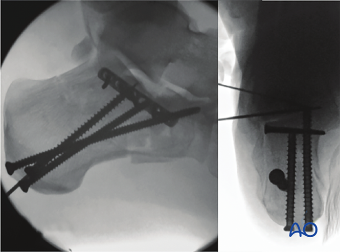
A rögzítéshez a K-drótokon túl percután csavarokat vagy lemezeket is használhatunk.
5. Műtét utáni kezelés
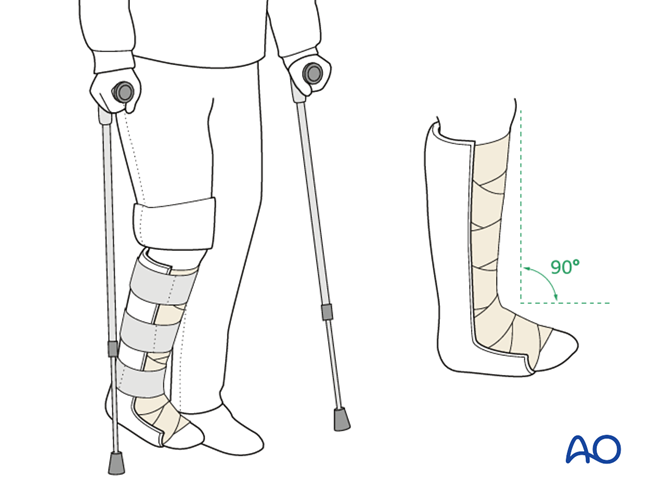
Kezdetben egy térdhajlati irányú, vastag merevítőt alkalmaznak az boka semleges helyzetének fenntartására, és a sérült végtagot időszakosan emelik.
A boka mozgásterjedelmére irányuló gyakorlatokat a műtét után 2–5 nappal kezdik el. A tűcsatorna-fertőzés megelőzése érdekében rendszeresen tisztítják a műtéti területet.
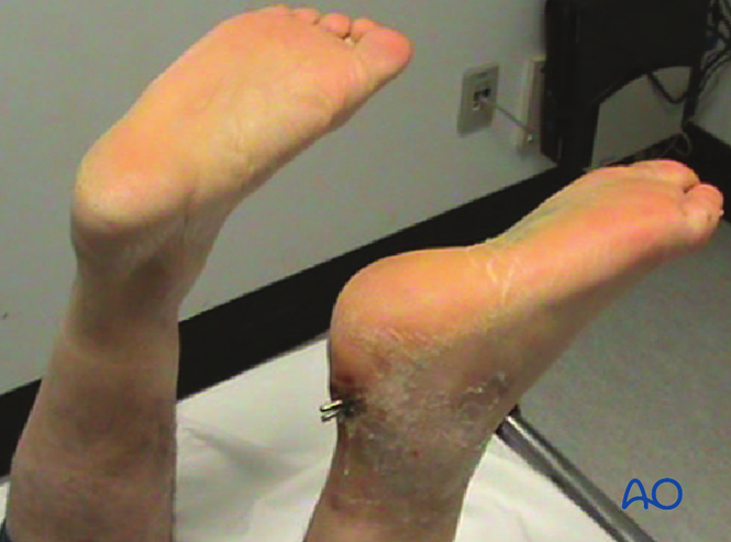
K-drótok eltávolítása: legkorábban 6 hetes korban (a példaképen a K-drót állapota 6 hetes időpontban látható).
Terhelés: csak a tűk eltávolítása után, 6 hetes korban kezdődik.
Képalkotásos követés: a műtét után 6, 12 és 26 héttel oldali és tengelyirányú röntgenfelvételeket készítenek.