EN
EN


Caution: These Are the Pitfalls in Internal Fixation Surgery for Proximal Humeral Fractures
I. Open Reduction and Internal Fixation (ORIF) for Proximal Humeral Fractures
1. Missed Greater and Lesser Tuberosity Fractures
Greater tuberosity (GT) fractures are often displaced posterosuperiorly and may appear minimally displaced on AP radiographs. Impacted lesser tuberosity (LT) fractures commonly occur with posterior shoulder dislocations. Obtain axillary and scapular Y (transscapular) radiographs and consider CT scans to accurately assess these fractures.
2. Ignoring Physiologic Age
Consider the patient’s physiologic age (activity level and functional demands) when making surgical decisions. A healthy, highly active elderly patient may benefit more from surgery than a younger, unhealthy, inactive patient.
3. Axillary Nerve Injury
The axillary nerve is at risk during both the deltopectoral and deltoid-split approaches. Identify and protect the nerve during dissection, retractor placement, and plate application to avoid injury.
4. Subacromial Impingement
When fixing GT fractures with screws, keep implants low on the tuberosity to avoid subacromial impingement with shoulder motion.
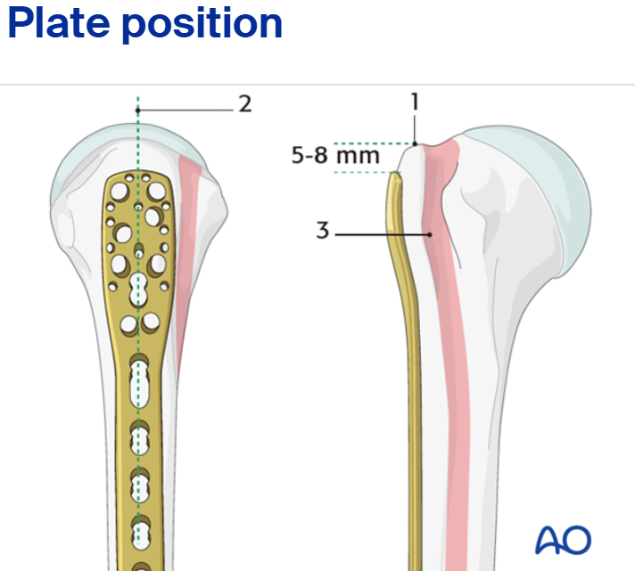
5. High Plate Placement
The most common error with plate fixation is placing the plate too high on the proximal humerus. This can cause subacromial impingement. Ensure the plate is positioned low enough to avoid this complication.
(AO Surgery Reference)
6. Drilling Through the Articular Surface
Do not drill through the articular surface of the humeral head. This increases the risk of screw penetration into the joint and can lead to varus collapse and arthritis.
7. Inadequate Tuberosity Fixation
Poor fixation of the tuberosities can lead to malunion, nonunion, and loss of shoulder function. Use a combination of screws, sutures, and/or tension-band constructs to achieve secure fixation, paying attention to bone quality.
II. Intramedullary Nailing (IMN) for Proximal Humeral Fractures
1. Cortical Breach During Canal Preparation
Breaching the cortex during canal preparation can lead to neurovascular injury or misplacement/migration of the nail. Use a gentle technique and follow the natural curvature of the humerus with awls and reamers.
2. Inadequate Distal Nail Insertion
Ensure the nail is inserted sufficiently into the distal fragment/medullary canal (at least 5 cm past the fracture) to provide adequate stability. Failure to do so can compromise fixation and lead to nonunion.
3. Prominent Nail Proximally
Avoid proximal prominence of the nail, as this can cause rotator cuff irritation and subacromial impingement. Ensure the nail is appropriately countersunk.
4. Misaligned/Errant Reaming
Reaming should be performed with the fracture anatomically reduced and aligned. Any varus or translational malalignment during reaming will result in a similar deformity when the nail is inserted.
5. Distal Cortex Penetration
Avoid penetrating the distal humeral cortex with the nail, especially in osteoporotic bone. This can cause an iatrogenic distal humeral fracture or pain. Choose the shorter of two nail lengths if unsure.
6. Screw Penetration
Avoid screw penetration through the subchondral bone/articular surface into the joint with locking screws. This can lead to humeral head collapse and screw protrusion into the joint.
7. Inadequate Proximal Screw Fixation
Use maximal (three to four) screw fixation rather than minimal screw fixation in the proximal humerus to provide better stability, particularly in osteoporotic bone.