EN
EN


X-ray interpretation (ABCS approach)
TheABCS approach of X-ray interpretation involves assessing the following:
- Alignment and joint space
- Bone texture
- Cortices
- Soft tissues
General points
Don’t forget to reviewall views, compare both sides and re‐examine any previous imaging.
If you spot one abnormality,do not lose focus until you have reviewed all areas of the image, otherwise, you might miss important pathology.
Alignment and joint space
Changes in alignment suggest afracture, subluxation (partial dislocation) or dislocation.
When describing the displacement, the position of thefragment distal to the fracture site is always described.
The radiograph below demonstrates why it is important to havemore than one view.
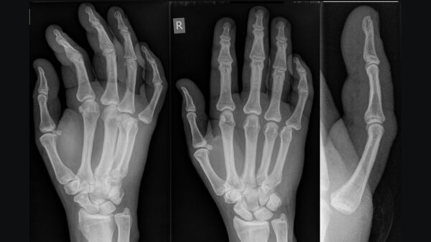
Joint dislocation of the metacarpophalangeal joint of the 5th digit.1
Carefully look at thejoint space to identify changes such as joint space narrowing due to cartilage loss, cartilage calcification (as in chondrocalcinosis) or new bone formation (e.g. osteophytes). Subchondral sclerosis (increased bone density) is often present along the joint lines in patients with osteoarthritis.
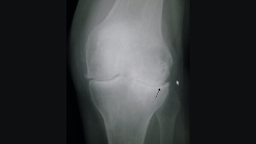
Osteoarthritis of the left knee. Note the osteophytes (right side of joint), narrowing of the joint space, and increased subchondral bone density (black arrow).2
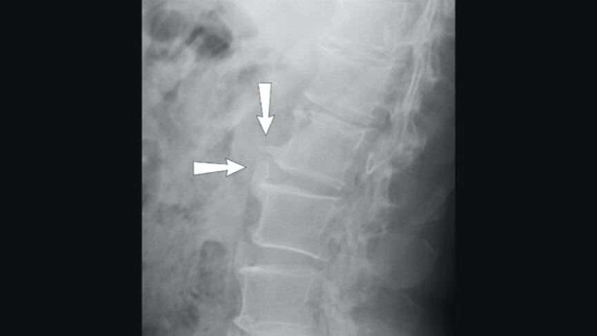
Lateral X-ray of spondylosis of the lumbar spine, with osteophytes marked by arrows3
Bone texture
Altered density or disruption in the usual internal matrix of fine white lines (trabeculae) within the substance of the bone and the thick external covering (cortex) may indicate pathology.
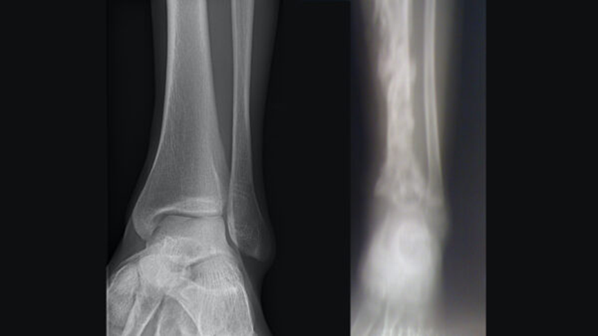
Healthy cortex and trabeculae vs osteomyelitis.4,5
Cortices
Trace around the outline of each bone as anystep in the cortex may indicate a fracture or other pathology.
Infection and tumours (primary and secondary) are the commonest causes of bony destruction.
A periosteal reaction is the formation of new bone in response to injury or other stimuli of the periosteum surrounding the bone. It may be the only sign visible to denote a problem with the bone (stress or healing fracture, mild osteomyelitis or tumour).
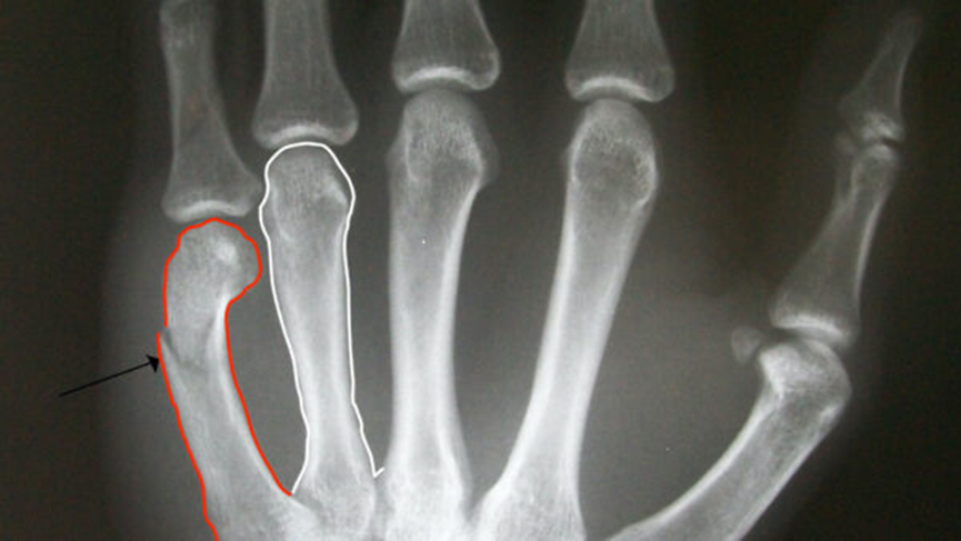
Trace around the bone cortex carefully to identify fractures (arrow)6
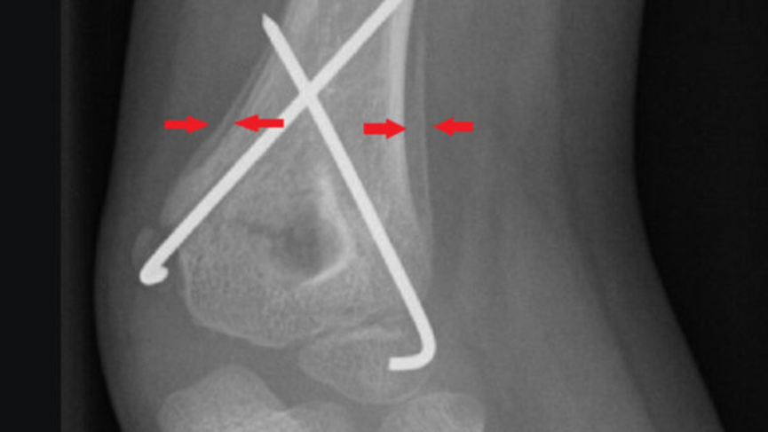
Periosteal reaction by a supracondylar fracture7
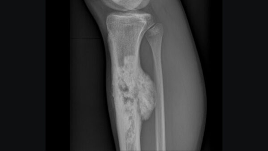
Osteosarcoma affecting the tibia8
Soft tissues
Look for anyswelling, foreign bodies or effusions. Sometimes soft tissue injuries are easier to visualise and can prompt a closer inspection for bony pathology.
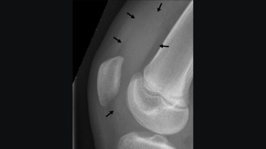
X-ray of the knee of a 12-year-old male, showing knee effusion of medium severity, marked by black arrows. It displaces the patella anteriorly and extends into the suprapatellar bursa.9
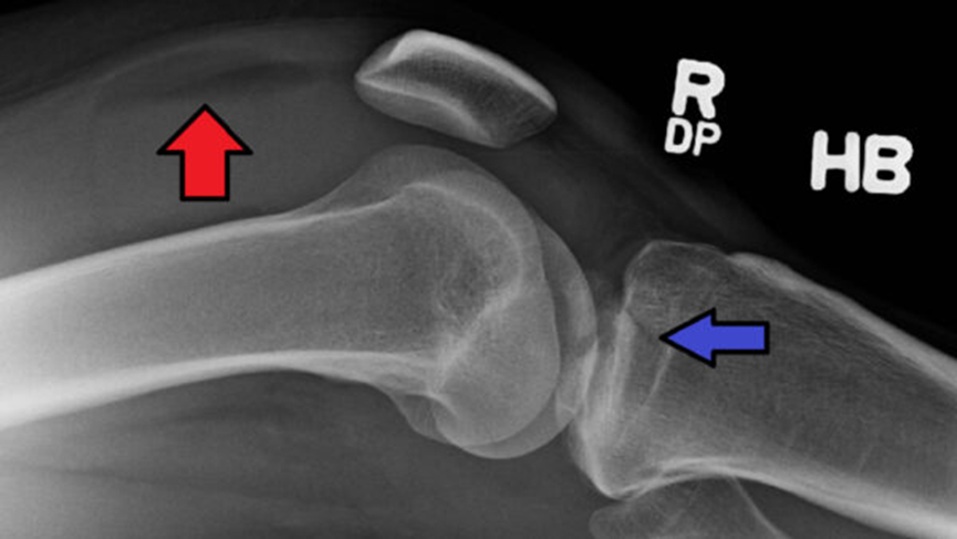
Lipohaemarthrosis (red arrow) due to a tibial plateau fracture (blue arrow)10