EN
EN


Femoral Neck Fracture Fixation Techniques: Latest Research Advances—A Narrative Review
Overview
Femoral neck fracture is a difficult problem that urgently needs to be overcome in orthopedic clinical practice; this type of fracture significantly increases patient disability and mortality rates. In young adults, femoral neck fractures are mostly caused by high-energy trauma and are often accompanied by multiple other injuries, making them a high-risk trauma type. In contrast, fractures in elderly patients are mostly low-energy injuries occurring on a background of osteoporosis, but due to the superimposition of underlying diseases, the risks of disability and death are often even higher. At present, clinical treatment of femoral neck fractures has reached a unified consensus: surgical treatment is the first choice. Surgical options are mainly divided into two major categories: internal fixation and arthroplasty. The femoral head has “three major” blood supplies: ① More than 70% of blood supply comes from the retinacular arteries arising from the medial and lateral circumflex femoral arteries; this is the most important blood supply line of the femoral head. ② About 20% comes from the nutrient artery of the femoral shaft. This artery enters the medullary cavity at the midshaft of the femur and can only reach the femoral neck, providing only a small amount of blood supply and contributing limitedly to the femoral head. ③ About 5% comes from the artery of the ligamentum teres, which supplies only the region around the fovea of the femoral head and has a weak blood supply role in adults. The degree of vascular injury is closely related to the degree of fracture displacement, and impaired blood supply directly increases the risk of osteonecrosis of the femoral head (ONFH). Therefore, when choosing internal fixation versus arthroplasty clinically, it is necessary to comprehensively evaluate indicators such as fracture displacement, bone stock defects, and patient age—all of these factors can increase the risk of complications such as fixation failure, nonunion, and malunion, severely impairing patients’ limb function and quality of life. Internal fixation is the foundational surgical procedure for femoral neck fractures. Over many years, various internal fixation techniques have been continuously developed and improved domestically and internationally, with the aim of achieving stable fixation of the fracture ends, promoting fracture healing, and maximally restoring affected-limb function. This review systematically summarizes various internal fixation strategies for femoral neck fractures and provides an in-depth explanation of indication selection, mainstream procedures, and frontier development directions for internal fixation surgery.
01 Selection Strategies for Internal Fixation
Before introducing various internal fixation procedures for femoral neck fractures, it is first necessary to clarify the indications for internal fixation surgery. Existing literature reports that the surgical failure rate and reoperation rate after internal fixation for femoral neck fractures range from 8% to 21%, with substantial individual variation. Therefore, fully understanding the various risk factors affecting internal fixation outcomes is crucial for clinical decision-making.
1.1 Fracture Displacement and Classification Systems
When determining the treatment plan for femoral neck fractures, the degree of fracture displacement is the primary assessment indicator, as displacement can directly predict the risk of vascular injury. Although multiple fracture classification systems are currently available for evaluating displacement, no single classification can completely guide treatment and predict prognosis. The most widely used in clinical practice remains the Garden classification. This classification was proposed by British orthopedic surgeon Robert Symon Garden in 1961. Based on fracture displacement, fracture completeness, and the alignment relationship of the trabeculae between the femoral head and neck, it divides femoral neck fractures into four types. Garden type I and II are nondisplaced fractures: type I is a valgus-impacted incomplete fracture; type II is a complete fracture without displacement. Most clinicians preferentially choose internal fixation for such fractures.
1.2 Pauwels Angle (Fracture Inclination Angle)
The Pauwels angle is used to describe the vertical inclination of the fracture line and is another core indicator guiding treatment. According to the direction of the fracture line, the Pauwels classification divides femoral neck fractures into three grades: grade I: Pauwels angle <30°; grade II: 30° ≤ Pauwels angle ≤50°; grade III: Pauwels angle >50°. In this classification system, the larger the Pauwels angle (the more vertical the fracture line), the more susceptible the fracture ends are to shear forces, the worse the stability, and the significantly higher the risk of internal fixation failure compared with horizontal fracture patterns. Therefore, for femoral neck fractures with a large Pauwels angle, poor internal fixation prognosis should be anticipated in advance.
1.3 Bone Quality and Age Factors
Initial post-injury displacement alone cannot completely determine internal fixation prognosis: some severely displaced fractures have good outcomes after internal fixation, whereas minimally displaced fractures may also experience internal fixation failure. Bone quality is another key influencing factor and is highly correlated with patient age. Although other conditions such as reduction quality are also important, advanced age combined with severe osteoporosis and extremely poor bone stock is an independent risk factor for internal fixation failure.
1.4 Fracture Posterior Tilt (Apex-Anterior Angulation)
Posterior tilt of the fracture (also called apex-anterior angulation) also affects internal fixation outcomes. The measurement definition of posterior tilt in the literature includes two types of changes: posterior angulation between the femoral head and neck and posterior displacement; both destroy fracture stability and injure nutrient vessels running through the fracture area. Palm et al. confirmed that posterior tilt ≥20° is the only independent risk factor predictive of reoperation. Although the Garden classification is simple to use and has been used for a long time, it has obvious shortcomings: interobserver agreement is very low, with kappa values of only 0.03–0.56, making it difficult to accurately reflect the degree of fracture posterior tilt. When relying solely on AP hip radiographs to classify fractures as Garden type I or II nondisplaced fractures and plan internal fixation, particular caution is needed; if the posterior tilt angle is significant, it must be fully corrected intraoperatively, and in some cases internal fixation should even be abandoned in favor of direct arthroplasty.
1.5 Surgical Timing
Surgical timing is an important factor determining internal fixation prognosis. There is general agreement in the field that surgery should be performed as early as possible, theoretically reducing the risk of post-traumatic femoral head necrosis and nonunion. A recent meta-analysis by Papakostidis et al. showed that the interval from injury to internal fixation surgery for femoral neck fracture was not statistically associated with the incidence of femoral head necrosis; however, surgery more than 24 hours after injury significantly increased the risk of nonunion (odds ratio [OR] = 0.33, P < 0.01).
1.6 Reduction Method: Open Reduction vs. Closed Reduction
Before surgery, the reduction strategy must be selected: open reduction or closed reduction. Compared with closed reduction, open reduction more readily achieves anatomic reduction but is more traumatic and is mostly used for markedly displaced fractures. In addition to internal fixation, displaced femoral neck fractures can also be treated with arthroplasty; therefore, in clinical internal fixation surgery, closed reduction is used more often than open reduction. Recent meta-analyses and randomized controlled trials have confirmed that open reduction and closed reduction show no significant differences in nonunion, femoral head necrosis, or overall complication rates. Patterson et al., based on a multicenter study from 12 level I trauma centers in North America, pointed out that for displaced femoral neck fractures in young and middle-aged patients, open reduction not only fails to significantly improve reduction quality but also increases the risk of reoperation; therefore, open reduction requires strict control of indications and should be used cautiously.
1.7 Length-Stable Fixation and Intraoperative Compression Concepts
There are two core concepts in femoral neck internal fixation: length-stable fixation and intraoperative compression fixation. The advantage of length-stable fixation is that it reduces postoperative femoral shortening; however, when the fracture is severely comminuted, the overall stability of this fixation method decreases, and the risks of nonunion and internal fixation failure increase. Related studies have also confirmed a higher reoperation rate in such cases. Therefore, for severely comminuted femoral neck fractures, blind use of length-stable fixation is not recommended.
1.8 Controversies in Implant Selection
There remains substantial controversy in clinical practice regarding the optimal choice of surgical implants for femoral neck fractures. Relevant clinical studies and biomechanical tests continue to be published, and novel implants and fixation techniques continue to be updated. The following section introduces current mainstream internal fixation implants and related latest research advances one by one.
02 Mainstream Internal Fixation Procedures for Femoral Neck Fractures
2.1 Dynamic Hip Screw (DHS)
The dynamic hip screw (DHS) is one of the most commonly used systems for internal fixation of femoral neck fractures. Its first-generation design was completed by Robert Danis in 1934, and it has a long history of application. The device was initially developed specifically for stable fixation of femoral neck fractures and was later continuously improved by scholars such as Ernst Pohl, Willis L. Pugh, and John Charnley, evolving into today’s mature modern DHS system. At present, its application scope is no longer limited to femoral neck fractures and covers fixation of various proximal femoral fractures (Figure 1).
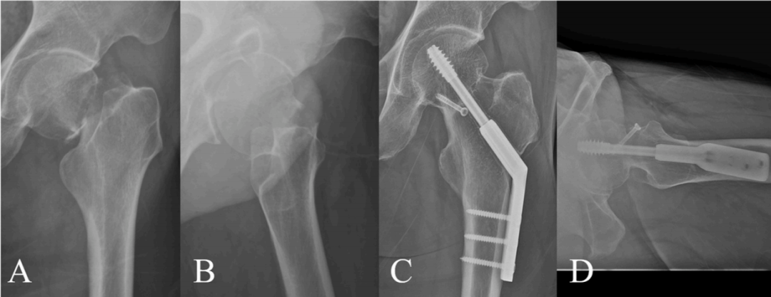
Figure 1. A 51-year-old man with an unstable femoral neck fracture treated with dynamic hip screw (DHS) internal fixation. Preoperative (A) AP and (B) lateral hip radiographs show a displaced femoral neck fracture; at 2-year postoperative follow-up, (C) AP and (D) lateral radiographs show no complications.
DHS is a core implant in the surgical instrument system for femoral neck fractures, and its design is based on two major mechanical principles: load sharing and controlled impaction. During surgery, a lag screw is passed across the fracture line and combined with a side plate attached to the femoral shaft to achieve axial compression and maintain fracture stability. DHS is especially suitable for minimally displaced stable fractures, with relatively ideal fracture union rates and affected-limb functional recovery. Clinically, DHS is often compared with multiple cannulated screw fixation: DHS requires a larger extent of soft tissue dissection and is associated with relatively more blood loss; however, DHS can provide stronger fixation stability. Deneka et al. conducted a biomechanical controlled study of internal fixation for unstable basicervical femoral neck fractures and confirmed that DHS fixation strength is superior to multiple cannulated screws. Clinical data from a recent systematic review and meta-analysis by Lim et al. showed that for high-angle Pauwels type II and III femoral neck fractures, the nonunion rate with DHS fixation was lower than that with multiple cannulated screw fixation (OR = 0.32; 95% confidence interval 0.11–0.96; P = 0.04), highlighting the therapeutic advantage of DHS in such fractures. There are preference differences in procedure selection among physicians in different regions. Through a cross-sectional survey combined with regression analysis, Bhandari et al. found that European surgeons were more likely than North American surgeons to prefer DHS over multiple cannulated screws. Both biomechanical and clinical studies have confirmed that DHS fixation strength is not inferior to, or is even superior to, multiple cannulated screws, with particularly prominent advantages in highly vertical fractures (Pauwels type II and III) and unstable fractures (Garden type III and IV).
2.2 Multiple Cannulated Screws (MCS)
Cannulated screw fixation is a minimally invasive alternative to DHS and is especially suitable for young and middle-aged patients with femoral neck fractures who have good bone stock and minimal displacement. The procedure relies on fluoroscopy or intraoperative imaging guidance to percutaneously place multiple screws across the fracture ends. Cannulated screws can protect femoral head blood supply, reduce surgery-related trauma, and shorten the recovery period (Figure 2).
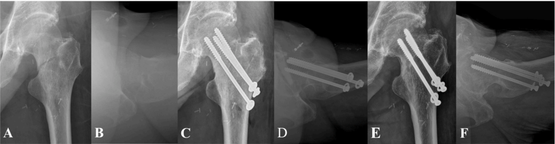
Figure 2. A 79-year-old man with a femoral neck fracture treated with multiple cannulated screw (MCS) internal fixation. Initial preoperative (A) AP and (B) lateral radiographs; immediate postoperative (C) AP and (D) lateral radiographs; final follow-up at 5 years postoperatively, (E) AP and (F) lateral radiographs.
After Garden, Barnes, and colleagues published related studies, multiple cannulated screw technique became widely popularized in the 1960s–1970s; by the mid-1980s, parallel screw placement gradually became mainstream, as this configuration allows moderate impaction at the fracture site. Biomechanical tests by Selvan et al. confirmed that triangular screw arrangement has greater resistance to failure than other arrangements. In the early 21st century, parallel and triangular screw configurations gained broad global acceptance. A recent meta-analysis by Lim showed that in multiple cannulated screw fixation for femoral neck fractures, parallel screw arrangement was not correlated with nonunion or postoperative ONFH. Multiple studies have used computer-aided design to explore optimal screw trajectories. In 2011, Filipov proposed the biplanar double-support screw placement technique to obtain additional cortical support. Clinically, an inverted triangular three-screw configuration is routinely used: the first screw is adjacent to the calcar, the second is near the posterior cortex, and the third is near the anterior cortex. Compared with DHS, cannulated screws are less traumatic, cause less intraoperative bleeding; in some cases, the procedure can be completely percutaneous without an incision, reducing soft tissue dissection, shortening hospital stay, and lowering treatment cost. However, for displaced femoral neck fractures, use of this procedure requires strict selection and attention to reduction quality. Kim et al. conducted a study of femoral neck fracture patients under 60 years of age: all 52 cases were treated with multiple cannulated screw fixation, and all were Garden type III or IV unstable fractures; the incidence of postoperative complications such as femoral head necrosis and nonunion was as high as 51.5%; in cases with complications, fracture posterior tilt on lateral radiographs was significantly greater (16.9° vs. 10.3°, P = 0.026).
2.3 Femoral Neck System (FNS)
The Femoral Neck System (FNS) is a newly developed device in recent years that integrates the dual mechanical advantages of compression and anti-rotation. FNS uses a screw-plate composite structure to achieve high-strength fixation; the blade combined with an anti-rotation screw design simultaneously improves axial stability and anti-rotation ability. Compared with DHS, FNS involves less soft tissue dissection and lower intraoperative bleeding risk; it is a minimally invasive fixation system that combines the advantages of both DHS and multiple cannulated screws. Multiple biomechanical studies have confirmed that FNS axial and rotational stability is comparable to conventional DHS and superior to multiple cannulated screws. Stoffel et al. showed that for unstable intracapsular femoral neck fractures (especially Pauwels type III), FNS biomechanical stability is equivalent to DHS and significantly superior to MCS; Moon et al., in an in vitro study of displaced basicervical femoral neck fractures, confirmed that even for extracapsular fractures, FNS fixation performance is not inferior to DHS. Clinical studies also support the advantages of FNS in treating adult femoral neck fractures. Patel et al. included 8 studies in a systematic review and meta-analysis: compared with traditional multiple cannulated screws, FNS was associated with lower postoperative complication rates and less femoral neck shortening, and better fracture union rates and affected-limb functional scores (all P < 0.001). Although existing in vitro tests and short-term clinical follow-up results are favorable, FNS has been on the market for a relatively short time and lacks long-term follow-up data; in addition, the device cost is relatively high and cost-effectiveness is insufficient. More studies are still needed to evaluate potential complications during implant removal. Yoon et al. reviewed 43 FNS surgical cases, of which 5 underwent implant removal; 3 had distal locking screw stripping, an incidence of 60%.
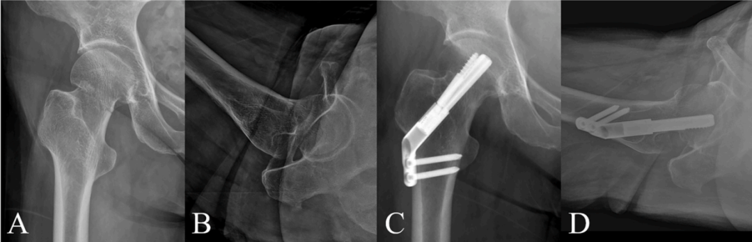
Figure 3. A 60-year-old woman with a stable femoral neck fracture treated with Femoral Neck System (FNS) internal fixation. Preoperative AP and lateral hip radiographs are shown in (A) and (B), respectively; at 1-year postoperative follow-up, (C) AP and (D) lateral radiographs show no complications.
2.4 Expanded Options: Modified Internal Fixation Procedures and Adjunctive Fixation Techniques
Intertrochanteric Valgus Osteotomy Combined With Internal Fixation
This procedure is a modified treatment for unstable femoral neck fractures, namely one-stage intertrochanteric valgus osteotomy combined with fracture internal fixation; it is mostly used for Garden type III and IV unstable fractures with vertically oriented fracture lines. During the same surgery as implant insertion, intertrochanteric valgus osteotomy is performed to alter the direction of mechanical forces across the vertical fracture surface, thereby reducing the risk of complications that occur frequently with this type of fracture.
Anteromedial Buttress Plate Fixation
Another type of adjunctive fixation technique is the anteromedial buttress plate, generally using a reconstruction plate to provide medial support to the femoral neck. In addition to conventional lateral fixation, this procedure adds plate support at the calcar, substantially improving overall stability; it is mostly used in young patients with adequate bone stock. Although related biomechanical and clinical studies exist, this strategy is not widely used clinically.
Conclusion
In the treatment of femoral neck fractures, internal fixation is a core treatment modality; clinical decisions require careful assessment, and it forms the foundation of the overall treatment strategy for femoral neck fractures. Current mainstream internal fixation procedures include multiple cannulated screws (MCS), dynamic hip screw (DHS), and Femoral Neck System (FNS). Based on existing biomechanical tests and clinical research evidence: when performing internal fixation for unstable femoral neck fractures, use of multiple cannulated screws requires particular caution, as its fixation strength is slightly inferior to that of dynamic hip screw and Femoral Neck System. As the latest-generation fixation device, Femoral Neck System has fixation strength comparable to dynamic hip screw while also offering the minimally invasive advantages of multiple cannulated screws; however, it should be objectively recognized that long-term follow-up data for this device are still lacking.
References:
1.Kim CH, Kim JW. A recent update on the fixation techniques for femoral neck fractures: A narrative review. J Clin Orthop Trauma. 2024 Jul 17;54:102497. doi: 10.1016/j.jcot.2024.102497. Erratum in: J Clin Orthop Trauma. 2026 Jan 08;73:103343. doi: 10.1016/j.jcot.2026.103343. PMID: 39157173; PMCID: PMC11326900.
2.Pearce JCH, Mitchell CJ. Fractures of the hip in younger adults. Surgery. 2023;41:200–206.
3.Handoll HH, Parker MJ. Conservative versus operative treatment for hip fractures in adults. Cochrane Database Syst Rev. 2008:CD000337.