
Get a Free Quote

Our representative will contact you soon.
Email
Name
WhatsApp
Company Name
Message
0/1000
locking compression plate lcp principles and clinical applications-0

News

Home >  News

Locking Compression Plate (LCP): Principles and Clinical Applications

Time : 2026-09-07

LCP Is Not a Panacea: Mechanical Boundaries and Pitfalls of Bridging Fixation

This review outlines the technical origins, biomechanical foundations, and clinical practice of the Locking Compression Plate (LCP). By utilizing combination holes, the LCP integrates two distinct internal fixation systems—absolute stability via conventional compression plating and relative stability via bridging plating—into a single implant. The authors note that most complications associated with the LCP are not due to inherent implant defects but rather to violations of core principles such as biological osteosynthesis, plate span, and screw density. The article collates in vitro experimental data and clinical case series to provide objective reference values for plate length, screw count, and screw configuration in different fracture scenarios.

Historical Evolution

Internal plate fixation has evolved continuously since Hansmann’s first procedures in 1886. The AO/ASIF standardized compression plating techniques, with the Dynamic Compression Plate (DCP) achieving axial compression at the fracture site through eccentric drilling, aiming for absolute stability and primary bone healing. However, this approach required extensive surgical exposure, stripping of soft tissues from bone fragments, leading to higher rates of infection, nonunion, and delayed union. In the 1980s, increasing complications with compression plating in diaphyseal fractures, alongside the success of intramedullary nailing which achieved secondary healing through callus formation, prompted new thinking. The concept of bridging plating was introduced, where surgery only corrects alignment, length, and rotation without disturbing the comminuted fracture zone. The plate spans the fracture segment and is anchored into the proximal and distal main fragments. This method provides only relative stability, allowing callus formation, and is also termed biological osteosynthesis.

Technical Core of the LCP

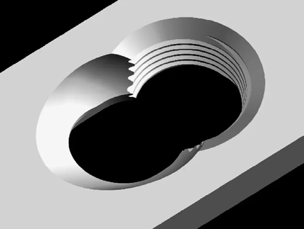
The Locking Compression Plate (LCP) builds upon the experience of PC‑Fix and LISS systems. Its revolutionary feature is the integration of two completely different fixation mechanisms within a single plate. The combination holes allow surgeons to choose either conventional dynamic compression, insertion of locking head screws to create an internal fixator, or a hybrid combination of both screw types (Figure 1).

Locking Compression Plate (LCP): Principles and Clinical Applications
Figure 1. The combination hole principle of the LCP, permitting the surgeon to apply both internal fixator and dynamic compression principles at the fracture site as needed.

Conventional plates rely on screws pressing the plate against bone, generating stability through plate‑to‑bone friction. When used as an internal fixator, the LCP does not require close contact with the cortical surface. However, this new technology imposes novel demands on surgical technique. The review emphasizes that a significant proportion of LCP‑related problems stem from neglect of the fundamental principles of biological bridging osteosynthesis.

Key Clinical Parameters for LCP Application

Plate Length

In conventional plating systems, shortening the plate reduces soft tissue dissection. This logic does not apply to the LCP. The LCP allows the use of longer plates without additional soft tissue trauma; plate selection should be governed solely by the fracture’s biomechanical status. The plate‑span ratio is defined as the total plate length divided by total fracture length. For comminuted fractures, this ratio should generally be 2‑3; for simple fractures, it should be 8‑10. Increasing the span reduces the load borne by the plate.

Screw Count and Configuration


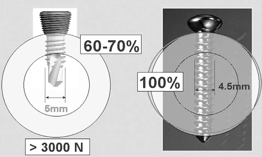
Screw density is defined as the number of screws inserted divided by the total number of plate holes; this value should be kept below 0.4‑0.5. Unlike conventional plates, the LCP does not mandate a fixed number of screws or cortices per bone fragment. At least two monocortical screws in each main fragment are recommended to maintain structural stability. For safety, 2‑3 screws per main fragment are generally placed, so that even if one screw has suboptimal purchase, overall stability is preserved. At least one bicortical screw per main fragment improves screw‑bone interface performance but does not reduce the probability of screw failure itself. Axial pullout strength is determined by the screw outer diameter; a 5.0 mm locking head screw (LHS) used monocortically achieves approximately 70% of the holding power of a 4.5 mm conventional bicortical screw (Figure 3).

Locking Compression Plate (LCP): Principles and Clinical Applications
Figure 3. Pullout test results: monocortical locking head screws achieve 70% of the holding power of conventional 4.5 mm bicortical screws.

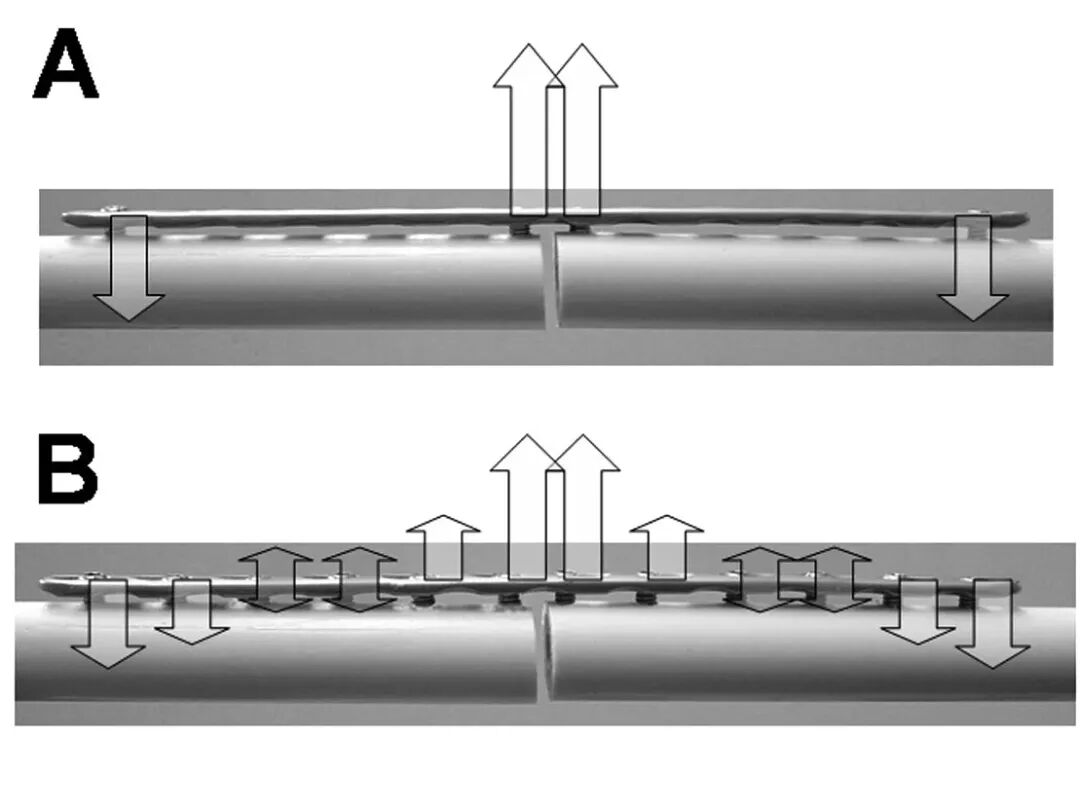
The number of screws alters the distribution of bending forces. The force distribution patterns differ significantly between minimum and maximum screw counts (Figure 2).

Locking Compression Plate (LCP): Principles and Clinical Applications
Figure 2. Force analysis corresponding to screw number: differences in bending force distribution between minimal (A) and maximal (B) screw configurations.

In comminuted fractures with no bone contact between main fragments, screws adjacent to the fracture zone should be inserted to increase the bridging length and disperse plate stress. In simple fractures with bone contact, 1‑2 combination holes on each side of the fracture should be left empty.

When drilling for locking head screws, axial deviation exceeding 5° significantly reduces structural stability; therefore, the aiming device must be used. When conventional and locking screws are used together, the conventional compression screws should be inserted first, followed by the locking head screws. Reversing this sequence may overload the screw‑bone interface.

Plate Contouring


When used as an internal fixator, the LCP does not require perfect bone contact. In diaphyseal fractures, gentle bending between screw holes allows screws to be directed in different orientations, providing multidirectional anchorage—particularly beneficial in osteoporotic bone. In metaphyseal regions, only gross contouring is needed to avoid excessive plate‑bone gaps and reduce soft tissue pressure, while allowing divergent screw placement to enhance overall pullout resistance.

Anatomically Pre‑contoured LCP Systems


Anatomically pre‑contoured LCPs no longer require extensive intraoperative bending; they assist in achieving anatomical reduction, and the accompanying aiming blocks facilitate accurate locking screw placement, standardizing surgical workflow. The review lists clinically available systems: PHILOS proximal humerus plate, distal humerus LCP, 2.4 mm distal radius system, hand‑specific LCP, distal femur LCP, proximal tibia LCP, distal tibia LCP, pilon‑specific LCP, medial tibial metaphyseal plate, condylar plate, and broad‑curved LCP. The broad‑curved LCP can accommodate the posterior curvature of the femur and is suitable for periprosthetic fractures and post‑knee arthrodesis fixation.

In Vitro Biomechanical Studies


Numerous in vitro biomechanical evaluations preceded clinical introduction. In a cadaveric model of distal radius C2 fractures, volar locking compression T‑plates demonstrated superior stability over conventional plates. In cadaveric radius specimens, the LCP outperformed LC‑DCP in anteroposterior stability and torsional resistance. In distal humerus fracture testing, the LCP achieved stable primary fixation. The PHILOS proximal humerus system, with its elastic properties, reduced peak stresses at the bone‑implant interface, resulting in lower early loosening rates in osteoporotic bone compared to stiffer implants. In long‑bone fracture models, the LCP exhibited higher yield strength than comparator implants. Some controlled studies did not show biomechanical superiority over conventional plates, but none demonstrated biomechanical inferiority of the LCP.

Clinical Outcomes


The LCP entered clinical use in 2001. Sommer (2003) published an early clinical series of 169 cases, concluding that the system had matured and offered particular value in complex fractures and revision settings after failed internal fixation. The distal radius is a high‑frequency application site. For dorsally displaced distal radius fractures, volar locking plates avoid dorsal extensor tendon irritation; the 2.4 mm LCP achieved good or excellent results in over 80% of osteoporotic distal radius fractures. Although direct implant costs are higher, the system permits early full range of active motion without loss of reduction. Literature reports document LCP use in sacral fractures, transverse sternal fractures, periprosthetic fractures, distal humerus fractures, and osteoporotic nonunions.

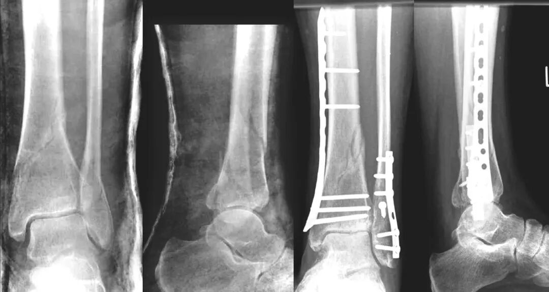
The review records that most LCP‑related complications are not material failures of the implant but rather failures to adhere to bridging osteosynthesis principles. Delayed union, though seen in some cases (notably in forearm fractures and open‑wedge proximal tibial osteotomies), occurred at a low overall rate. Figure 4 shows a case of distal tibial fracture managed with MIPO bridging fixation.

Locking Compression Plate (LCP): Principles and Clinical Applications
Figure 4. AO43‑A2 tibial shaft fracture: distal tibia LCP case; the fibula was fixed with conventional plating principles, while the tibia was fixed using MIPO bridging osteosynthesis.

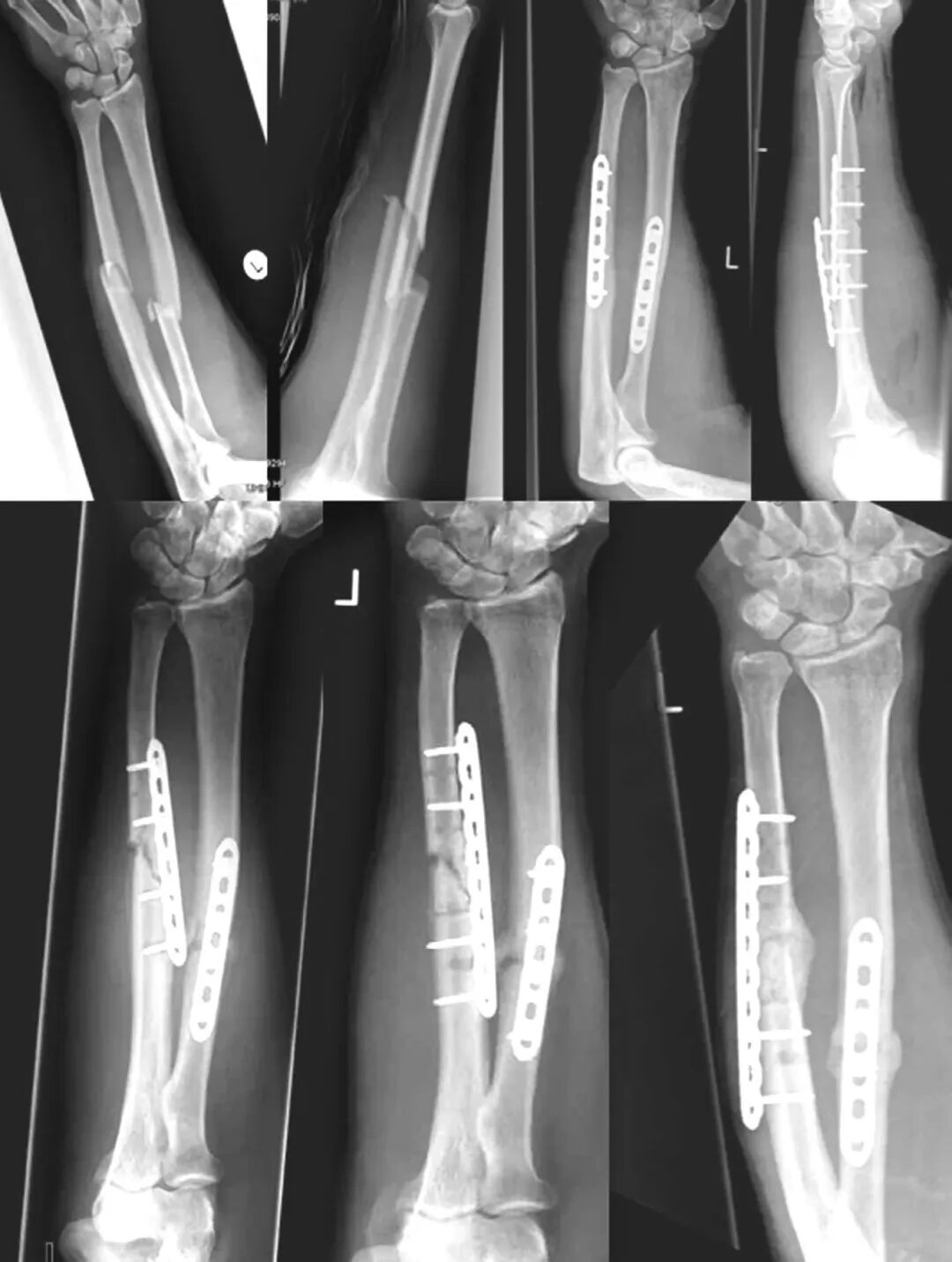
Failures due to inadequate bridging principles can be revised by using a longer plate and adjusting screw configuration. Figure 5 illustrates an ulnar shaft fracture where initial screws were placed too close to the fracture zone, leading to nonunion; after revision with a longer 10‑hole plate and repositioning of medial screws to increase bridging span, union was achieved at 3 months post‑revision.

Locking Compression Plate (LCP): Principles and Clinical Applications
Figure 5. AO22‑A3 forearm fracture LCP case: initial 8‑hole plate with screws too close to the fracture, resulting in nonunion at 5 months; revision with a longer 10‑hole plate, screw repositioning, and increased bridging length led to radiographic union at 3 months.

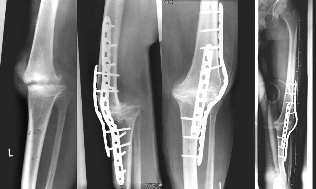
The LCP has also been used in arthrodesis revision. Figure 6 shows a case of knee arthrodesis nonunion after removal of an infected revision knee prosthesis and external fixation. A bent 20‑hole 4.5/5.0 mm LCP combined with another 10‑hole LCP and autogenous cancellous bone graft achieved fusion at 6 months postoperatively.

Locking Compression Plate (LCP): Principles and Clinical Applications
Figure 6. Chronic infection with nonunion after knee arthrodesis using external fixation; revision with a bent 20‑hole 4.5/5.0 mm LCP plus a 10‑hole LCP and autogenous cancellous bone graft, resulting in fusion at 6 months.

References (as derived from the review content)

  1. NIEMEYER P, SÜDKAMP N P. Principles and Clinical Application of the Locking Compression Plate (LCP). ACTA CHIRURGIAE ORTHOPAEDICAE ET TRAUMATOLOGIAE ČECHOSL., 2006,73:221‑228.
  2. BLATTER, G., WEBER, B. G.: Wave plate osteosynthesis as a salvage procedure. Arch. orthop. traum Surg., 109: 330‑3, 1990.
  3. FLORIN, M., ARZDORF, M., LINKE, B., AUER, J. A.: Assessment of stiffness and strength of 4 different implants available for equine fracture treatment: a study on a 20 degrees oblique longbone fracture model using a bone substitute. Vet. Surg., 34: 231–8, 2005.
  4. FRANKIE, L., XIANG, Z.: Locking compression plate fixation for periprosthetic femoral fracture. Zhongguo Xiu Fu Chong Jian Wai Ke Za Zhi, 16: 123–5, 2002.
  5. FRIGG, R.: Development of the Locking Compression Plate. Injury, 34 Suppl. 2: B6‑10, 2003.
  6. GARDNER, M. J., BROPHY, R. H., CAMPBELL, D., MAHAJAN, A., WRIGHT, T. M., HELFET, D. L., LORICH, D. G.: The mechanical behavior of locking compression plates compared with dynamic compression plates in a cadaver radius model. J. orthop. Trauma, 19: 597–603, 2005.

PREV : None

NEXT : Shanghai CareFix Medical Presents Orthopedic Solutions at the 7th World Congress of ASAMI-BR & ILLRS Societies

logo