EN
EN


Complex Pilon Fractures: The "Four Principles" and the "Four-Step Foundation Method"
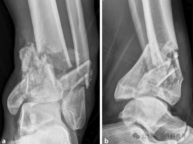
Pilon fractures were first reported in 1911 and are commonly caused by high-energy axial trauma. They account for approximately 5%–7% of tibial fractures and are often associated with fibular fractures. They typically present with comminution of the metaphysis and distal tibial articular surface, accompanied by severe soft tissue injury. The goal of Pilon fracture treatment is to restore normal congruity of the tibiotalar articular surface and prevent post-traumatic arthritis.
PART 1
The “Four Principles” of Pilon Fracture Fixation
Pilon fracture surgery is technically demanding. Severely comminuted fractures lack reduction references, and the tibiotalar articular surface has high reduction requirements, making Pilon fractures known as “the most challenging fractures.” In their publications in 1969 and 1973, Rüedi and Allgöwer summarized four major principles of internal fixation for Pilon fractures:
- Restoration of fibular length;
- Restoration of the smoothness/congruity of the distal tibial articular surface;
- Bone grafting of the tibial metaphysis;
- Medial tibial plate fixation.
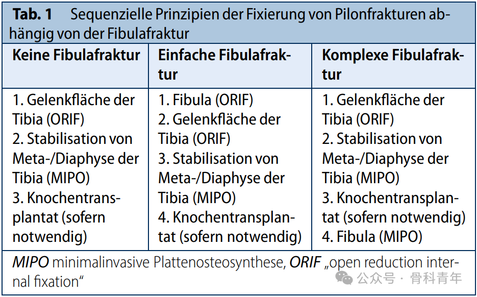
Guided by these principles, the stepwise principles for Pilon fracture reduction and fixation based on the fibular fracture status remain the clinical gold standard.
I. Fixation of Fibular Fractures
When treating fibular fractures, the key is to distinguish whether the fracture is simple or complex. For simple fibular fractures, open reduction and internal fixation (ORIF) with a plate is preferred. Following the principle of “fix the fibula first,” restoring and maintaining the normal length, alignment, and rotation of the fibula is crucial for subsequent reduction of the tibial articular surface.
If the fibula has a complex comminuted fracture that is difficult to reduce anatomically, it is recommended to begin with tibial reduction and fixation. After reduction and fixation of the tibial Pilon fracture are completed, the fibula is fixed using a bridging fixation technique, either ORIF or minimally invasive plate osteosynthesis (MIPO); this is the final step of the procedure.
II. Reduction and Fixation of the Tibial Articular Surface
To achieve good reduction of the tibial articular surface, the fracture pattern of the articular region and the position of the talus must first be precisely analyzed. For articular fractures with multiple fragments and impaction, the relatively intact talar articular surface can be used as a reference and support. Using the talus as a template, the tibial articular surface is reduced progressively from the center to the periphery. Before this step, it is essential to ensure that the talus is at the correct length relative to the reduced or intact fibula and is centered beneath the tibial shaft. Otherwise, malreduction of the tibial articular surface is likely.
For type C fractures, individualized articular reduction strategies should be developed according to the specific fracture morphology. In general, two main surgical strategies are available:
Option 1: Convert a type C fracture into a type B fracture.
If there is a large weight-bearing articular fragment that can extend directly and simply into the intact metaphyseal/diaphyseal region, the best approach for a type C fracture is to first reduce this weight-bearing articular fragment and fix it to the intact shaft, thereby converting the type C fracture into a type B fracture. After this, the remaining articular surface is anatomically reduced, and an appropriate implant is used to fix the entire fracture construct.
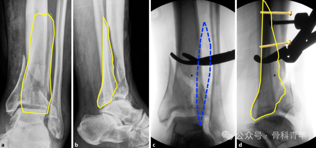
Figure a, b. Preoperative X-rays show a Pilon fracture (AO 43-C3). A large posterior articular fragment (yellow outline) can extend simply into the diaphyseal region; c, d. Through an anterolateral approach (c, blue dashed line), reduction forceps are used to anatomically reduce the posterior articular fragment (d, yellow outline) to the shaft, and it is fixed with two screws. This maneuver converts the type C fracture into a type B fracture.
Option 2: Convert a type C fracture into a type A fracture.
If the above strategy of converting a type C fracture into a type B fracture cannot be performed, the type C fracture can be converted into a type A fracture by reducing and temporarily fixing the articular block. When reducing the articular surface, the talus should be used as a template, and reduction should proceed from the center to the periphery. After articular surface reduction is completed, the metaphyseal fracture is addressed. The anatomically reduced articular block is temporarily fixed, then aligned with the shaft according to correct alignment, length, and rotation, and fixed with an appropriate implant. This method is particularly suitable for cases with a complex comminuted metaphyseal region and no clear bony reference between the articular block and the metaphysis/diaphysis.
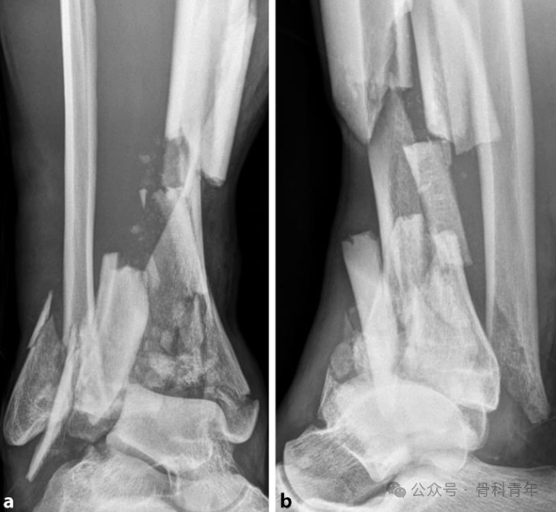
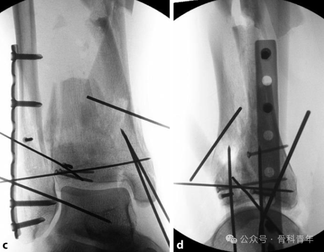
Figure a, b. Preoperative X-rays show a Pilon fracture (AO 43-C3). Because of the comminuted metaphyseal region, there is no reference between the articular fracture block and the intact shaft; c, d. Through an “extended lateral approach” combined with a small medial approach, the articular block is first anatomically reduced, thereby converting the type C fracture into a type A fracture. The fracture is then functionally aligned and fixed to the shaft.
III. Bone Grafting
In tibial Pilon fractures caused by high-energy injury, even after successful articular surface reduction, a large bone defect often remains in the metaphysis. Bone grafting is an effective adjunctive treatment. It not only provides additional support for the articular fracture fragments but also effectively prevents secondary displacement of the fragments postoperatively. For bone graft material, autologous cancellous bone or autologous bone chips are preferred, usually harvested from the patient’s ipsilateral iliac crest or ipsilateral proximal tibia. Autologous bone is chosen mainly because it has the best osteoinductivity and osteoconductivity and can better promote bone healing.
IV. Medial Buttress Plate Fixation
To counteract the varus deformity commonly seen in tibial Pilon fractures, Rüedi and Allgöwer proposed medial buttress plate fixation. In fact, accurately distinguishing the tension and compression forces generated by the trauma mechanism is crucial. Over the past few decades, the advent of locking compression plates (LCP) has led many orthopedic surgeons to no longer use medial buttress plates as the primary fixation support. Currently, anatomically precontoured angle-stable plates suitable for the medial, anterolateral, and posterior bone columns are available. In current clinical practice, surgeons flexibly select primary implants and other smaller auxiliary implants based on soft tissue conditions and the extent of bony exposure required. Medial plates can usually be placed subcutaneously through minimally invasive plate osteosynthesis (MIPO). This placement protects the soft tissues and also preserves sufficient space for a possible second, larger anterolateral or posterior primary surgical approach. Through rational selection and placement of implants, “biological internal fixation” is ultimately achieved, ensuring sufficient stability of the injured bone column.
PART 2
The Four-Step Foundation Method
Conventional internal fixation for Pilon fractures requires large incisions and extensive soft tissue dissection, and articular surface reduction cannot be well controlled. In recent years, the “Four-Step Foundation Method” for Pilon fractures proposed by Professor Li Dong’s team at the Second Hospital of Shanxi Medical University aims to accurately reduce the articular surface while reducing surgical trauma and has achieved good clinical results. The article was recently published in the JOSR journal and is shared for reference by orthopedic surgeons:
I. Surgical Incision
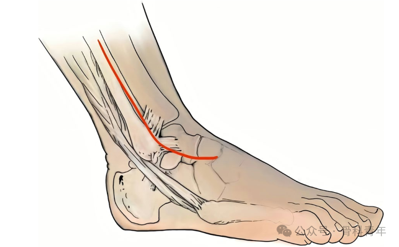
Unlike the classic widely used anteromedial approach, this method uses a long anterolateral incision, extending from the anterior border of the fibula through the lateral malleolus to the base of the fourth metatarsal, approximately 15 cm in length.
II. Reduction and Fixation of the Lateral Malleolus
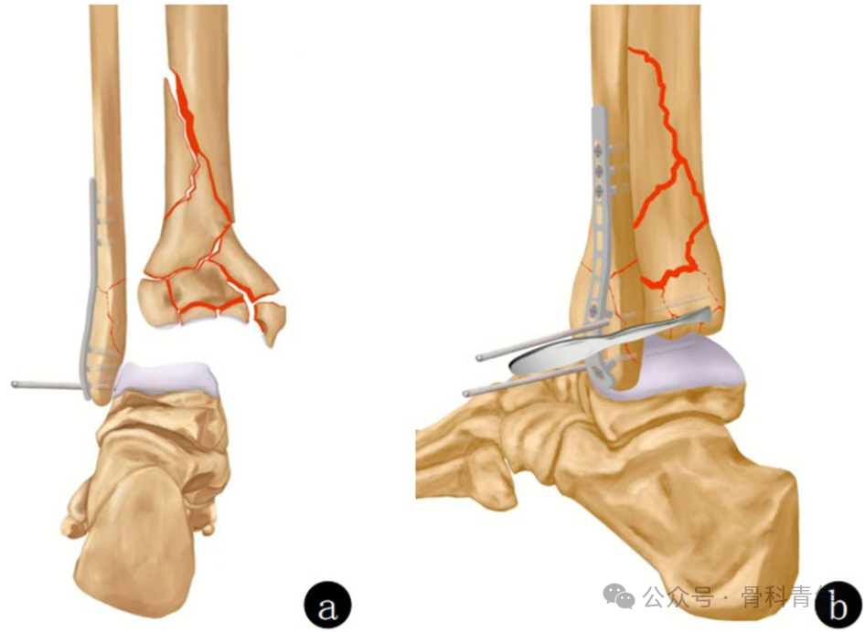
Restore the length and anatomical position of the lateral malleolus, and place an anatomical plate. The talus often displaces anterosuperiorly; press it downward so that its cartilage surface is flush with the medial side of the lateral malleolus. Using the normal “Mercedes-Benz triangle” of the tibia, fibula, and talus as a reference, maintain reduction with reduction forceps. Temporarily fix the lateral malleolus and talus with 2.0 mm Kirschner wires, avoiding the plate, and use the talus as a template to reconstruct the distal tibial articular surface.
III. Reduction and Fixation of the Posterior Malleolus
There are two methods for posterior malleolus fixation:
Method 1: Use a narrow periosteal elevator to pry from behind the posterior malleolus so that the posterior malleolar articular surface matches the talar articular surface;
Method 2: Manually reduce the posterior malleolus with fingers from the lateral side of the peroneus longus and brevis muscles.
After reduction of the posterior malleolus, fix it with a 1.5 mm Kirschner wire through the fibula.
IV. Reduction and Fixation of the Medial Malleolus
Make two small auxiliary incisions on the medial side of the ankle. From the distal incision, reduce the displaced medial malleolus with towel clips, assess the matching degree between the medial articular surface of the medial malleolus and the talar dome, temporarily fix with Kirschner wires, and then fix with a pre-contoured reconstruction plate. The most distal screw is inserted at a 45° angle downward, and the upper two screws are inserted at a 45° angle upward.
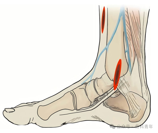
If the soft tissue conditions of the medial malleolus are poor and the risk of infection with plate placement is high, the medial malleolus can be fixed with Kirschner wires.
V. Reconstruction of the Distal Tibial Articular Surface
Follow the principle of “posterior to anterior, lateral to medial,” use the talar articular surface as a template for reduction, temporarily fix with Kirschner wires, and then fix with an L-shaped plate anterior to the ankle. Depending on the degree of fracture comminution and the status of plate fixation, determine whether to remove the Kirschner wires.
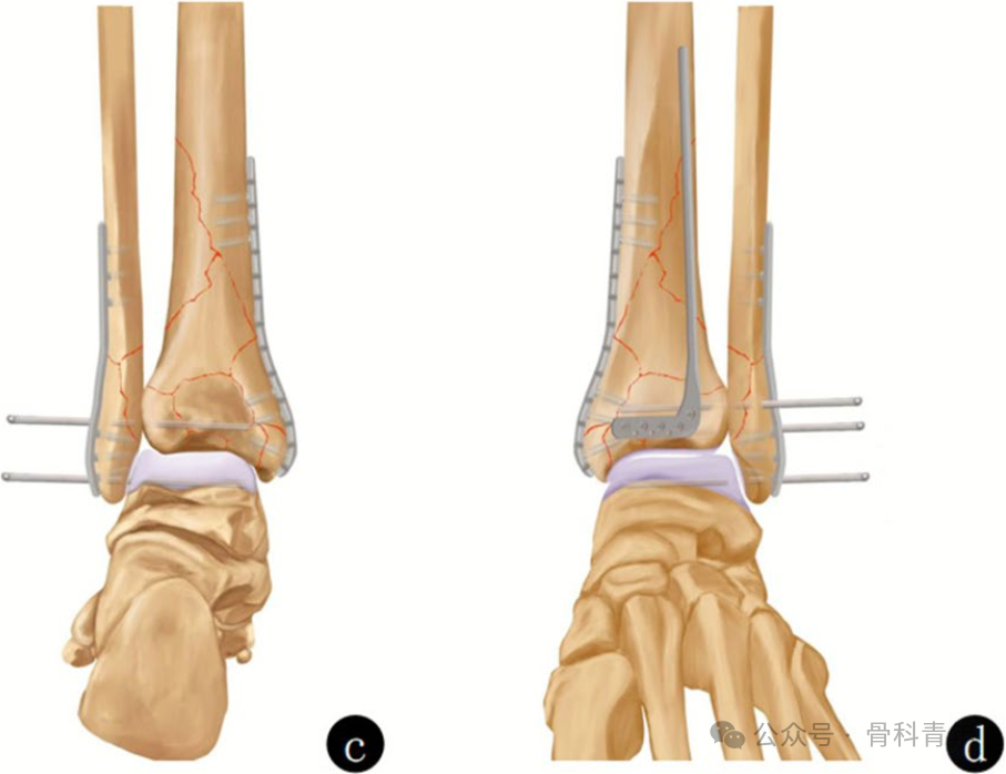
Both conventional single-incision and multiple-incision approaches for Pilon fractures have their respective drawbacks. Single-incision approaches have problems with exposure and soft tissue retraction; multiple-incision approaches have problems with soft tissue coverage and inter-incision spacing. The surgical method proposed by this team, using an anterolateral approach combined with a medial minimally invasive approach, is relatively convenient for both surgical exposure and reduction/fixation, and has significant clinical guiding significance for Rüedi–Allgöwer type III Pilon fractures.