Interbody Fusion Cage: Advanced Spinal Solutions

Get a Free Quote

Our representative will contact you soon.
Email
Name
Company Name
Message
0/1000

interbody fusion cage

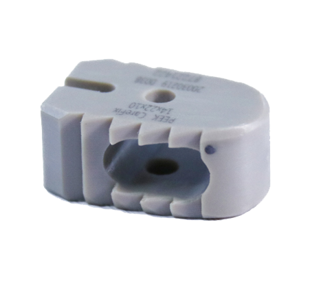
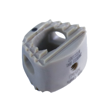
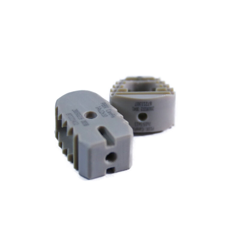
An interbody fusion cage is a medical implant designed to facilitate spinal fusion procedures by providing structural support between vertebral bodies. This specialized device is inserted into the intervertebral disc space after disc material removal, creating a stable environment that promotes bone growth and fusion. The interbody fusion cage serves as a scaffold that maintains proper disc height, restores spinal alignment, and decompresses neural structures while the fusion process develops. Manufactured from biocompatible materials such as titanium alloys, polyetheretherketone, or carbon fiber composites, these cages feature porous surfaces or openings that allow bone graft material to pass through, encouraging biological integration. The technological design incorporates anatomically contoured shapes that match the natural curvature of the spine, ensuring optimal fit and stability. Advanced engineering enables the interbody fusion cage to distribute mechanical loads evenly across the vertebral endplates, reducing stress concentration and subsidence risk. Various cage designs accommodate different surgical approaches, including anterior lumbar interbody fusion, posterior lumbar interbody fusion, transforaminal lumbar interbody fusion, and lateral lumbar interbody fusion techniques. Clinical applications span degenerative disc disease, spondylolisthesis, spinal stenosis, and trauma cases requiring stabilization. The interbody fusion cage represents a cornerstone technology in modern spine surgery, combining biomechanical stability with biological fusion promotion to achieve lasting therapeutic outcomes for patients suffering from debilitating spinal conditions.

New Product Recommendations

The interbody fusion cage delivers multiple practical benefits that directly impact surgical outcomes and patient recovery. First, these devices restore and maintain the natural height between vertebrae, which alleviates pressure on nerve roots and reduces pain symptoms immediately following surgery. This height restoration also corrects spinal alignment, improving posture and reducing mechanical stress on adjacent spinal segments. The structural support provided by the interbody fusion cage creates immediate stability, allowing patients to mobilize earlier in their recovery process compared to fusion procedures without cage support. The open architecture design facilitates bone graft incorporation, significantly improving fusion rates by allowing direct contact between the graft material and the vertebral endplates. This biological integration creates a permanent bony bridge that eliminates motion at the affected segment, addressing the root cause of pain. From an operational perspective, surgeons benefit from standardized sizing options and instrumentation that streamline surgical workflows and reduce procedure time. The radiolucent properties of certain cage materials enable clear postoperative imaging, allowing physicians to monitor fusion progress accurately without metal artifacts obscuring the view. The interbody fusion cage suits diverse patient populations and pathologies, from young active individuals to elderly patients with osteoporotic bone, thanks to varied design options and surface treatments. Cost-effectiveness emerges through reduced revision surgery rates, as the enhanced stability and fusion promotion minimize implant failure risks. Patients experience improved quality of life through pain reduction, restored function, and the confidence that comes with a proven surgical solution. The decision to use an interbody fusion cage represents an investment in long-term spinal health, backed by extensive clinical evidence demonstrating superior fusion rates and patient satisfaction compared to alternative approaches.

Practical Tips

Cannulated Screw Types: Features and Benefits Explained

16

Dec

Cannulated Screw Types: Features and Benefits Explained

Orthopedic surgery has evolved significantly over the past decades, with advances in implant technology playing a crucial role in improving patient outcomes. Among these innovations, the cannulated screw stands out as a versatile and highly effective...
View More
Application of carbon fiber material external fixation bracket in clinical practice

13

Jan

Application of carbon fiber material external fixation bracket in clinical practice

The integration of advanced materials in orthopedic surgery has revolutionized patient care and treatment outcomes. Among these innovations, carbon fiber external fixation systems have emerged as a superior alternative to traditional metal frameworks...
View More
Evolution of Intervertebral Fusion Device Materials: Clinical Efficacy Comparison between PEEK and Titanium Alloy

13

Jan

Evolution of Intervertebral Fusion Device Materials: Clinical Efficacy Comparison between PEEK and Titanium Alloy

The advancement of spinal surgery has been significantly influenced by the development of sophisticated intervertebral fusion device technologies. Modern spine surgeons rely heavily on these devices to achieve successful fusion outcomes while minimiz...
View More
Taylor brace: a new choice for minimally invasive treatment of complex fracture repair

11

Feb

Taylor brace: a new choice for minimally invasive treatment of complex fracture repair

The Taylor brace represents a significant advancement in orthopedic trauma management, offering surgeons and patients a minimally invasive alternative for treating complex fracture repairs. This innovative external fixation system has transformed the...
View More

Get a Free Quote

Our representative will contact you soon.
Email
Name
Company Name
Message
0/1000

interbody fusion cage

Advanced Biomechanical Stability

Advanced Biomechanical Stability

The interbody fusion cage provides exceptional biomechanical stability through its engineered design that optimizes load distribution across the vertebral endplates. Unlike traditional fusion techniques that rely solely on posterior fixation, the cage placement within the anterior column of the spine harnesses the natural compressive forces of body weight to enhance stability. This anterior column support reduces stress on posterior instrumentation, decreasing the risk of hardware failure. The cage geometry features teeth, ridges, or textured surfaces that grip the endplate bone, preventing migration and subsidence during the critical healing period. Material selection plays a crucial role, with modulus of elasticity matched to bone properties to minimize stress shielding while providing adequate support. The three-dimensional structure resists rotational, flexion, and extension forces, creating an environment where motion at the fusion site is minimized. This mechanical stability is essential for successful bone healing, as excessive micromotion can inhibit osteogenesis. Patients benefit from earlier mobilization and reduced reliance on external bracing, accelerating return to daily activities and improving overall satisfaction with surgical outcomes.
Enhanced Biological Fusion Promotion

Enhanced Biological Fusion Promotion

The interbody fusion cage functions as a biological scaffold that actively promotes bone fusion through multiple mechanisms. The device features strategically placed windows, perforations, or porous surfaces that allow packed bone graft material to interface directly with the bleeding bone of the vertebral endplates. This direct contact is essential for osteoinductive and osteoconductive processes that drive new bone formation. The cage protects the graft material from compression and displacement, maintaining graft volume and position throughout the fusion process. Surface treatments such as plasma spray coatings, roughened textures, or bioactive materials further enhance osseointegration by encouraging cellular attachment and proliferation. The interbody fusion cage design maintains the intervertebral space at the optimal height, ensuring proper tension on the surrounding ligaments and annulus, which contributes to stability and vascular ingrowth. The contained environment within the cage creates an ideal biological chamber where growth factors, cells, and matrix can interact without interference. Clinical studies consistently demonstrate higher fusion rates with interbody fusion cage usage compared to cage-free techniques, translating to better long-term outcomes and reduced need for revision surgery.
Versatile Surgical Application

Versatile Surgical Application

The interbody fusion cage offers remarkable versatility across multiple surgical approaches and patient presentations, making it an indispensable tool in modern spine surgery. Surgeons can select from various cage footprints, heights, and lordotic angles to match individual patient anatomy and pathology precisely. This customization ensures optimal correction of deformity and restoration of sagittal balance, which are critical factors in surgical success. The device accommodates anterior, posterior, lateral, and oblique surgical corridors, allowing the surgeon to choose the approach that minimizes tissue disruption while maximizing access to the pathology. For revision surgeries where previous hardware or scarring complicates access, alternative approach options with interbody fusion cage placement provide viable solutions. The cage material options, including titanium for strength, polyetheretherketone for radiolucency, or composite materials for specific mechanical properties, allow selection based on imaging requirements and patient factors such as bone quality or allergies. Expandable cage designs accommodate minimal access surgery techniques, reducing incision size and muscle trauma. The interbody fusion cage adapts to single-level or multi-level fusion constructs, standalone applications or supplemented with posterior fixation, providing flexibility to address simple to complex spinal pathologies with a consistent, proven technology platform.
logo