Distal Tibia External Fixator Solutions

Get a Free Quote

Our representative will contact you soon.
Email
Name
Company Name
Message
0/1000

distal tibia external fixator

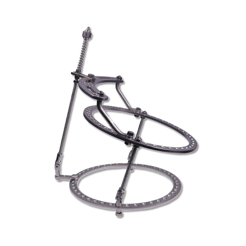
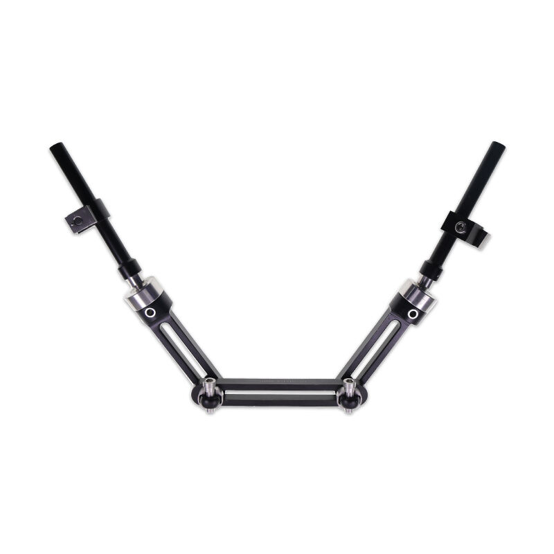
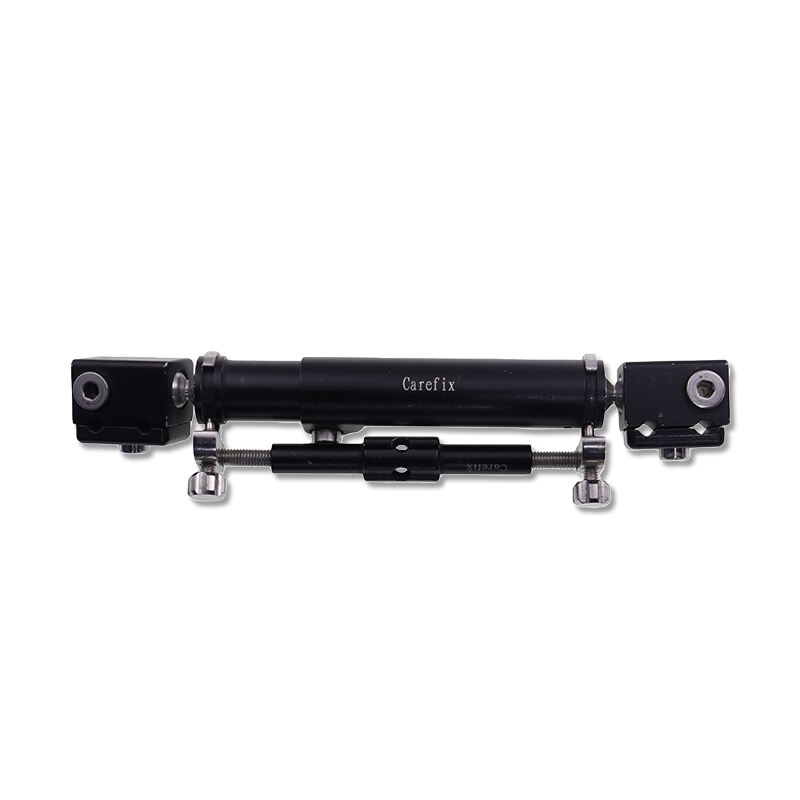
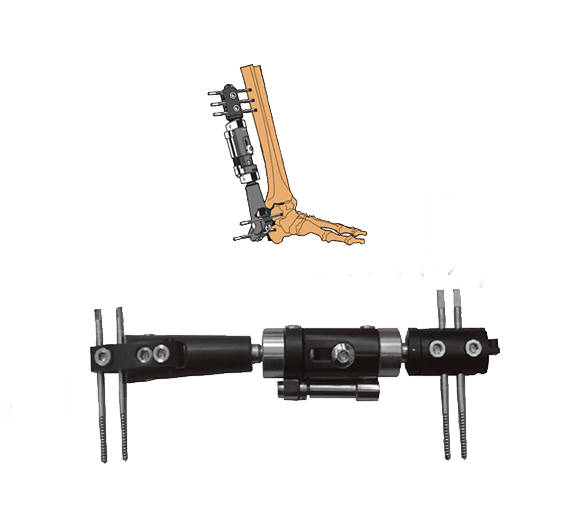
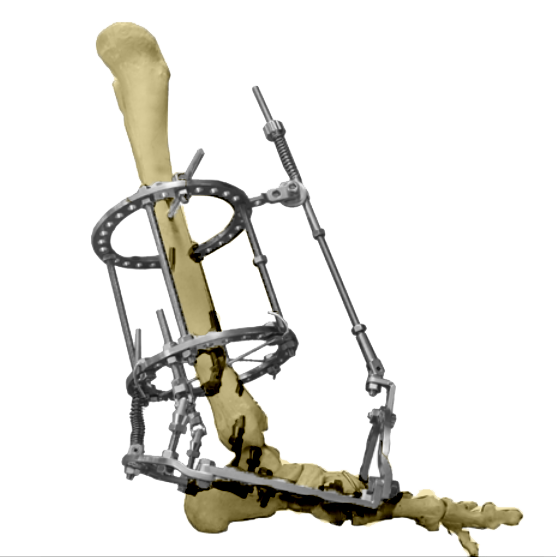
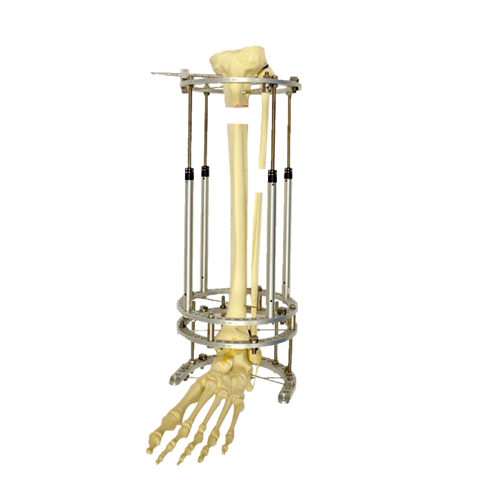
The distal tibia external fixator is a specialized orthopedic device designed to stabilize and treat fractures, deformities, and injuries occurring in the lower leg region near the ankle. This medical apparatus consists of metal pins or wires that penetrate the bone and connect to an external frame, providing rigid support while allowing the injured bone to heal properly. The distal tibia external fixator serves as a critical solution for complex lower leg injuries where traditional internal fixation methods may not be suitable. Its main functions include maintaining bone alignment, controlling limb length, correcting angular deformities, and facilitating soft tissue management during the healing process. The technological features of this device incorporate adjustable components that enable precise three-dimensional control of bone fragments, allowing surgeons to make gradual corrections throughout the treatment period. The modular design accommodates various anatomical configurations and injury patterns, making it adaptable to different patient needs. Modern versions incorporate radiolucent materials that permit clear visualization during X-ray imaging without interference from the fixation apparatus. Applications of the distal tibia external fixator extend to traumatic fractures, including open fractures with significant soft tissue damage, comminuted fractures with multiple bone fragments, infected nonunions requiring stabilization, limb lengthening procedures, and correction of post-traumatic deformities. This device proves particularly valuable in emergency settings where immediate stabilization is necessary before definitive surgical intervention can be performed.

New Product Releases

The distal tibia external fixator offers numerous practical benefits that directly address the challenges faced by orthopedic surgeons and their patients. One significant advantage is the minimally invasive nature of the procedure, which reduces surgical trauma to already compromised soft tissues surrounding the injury site. Unlike internal fixation methods that require extensive surgical exposure, this external approach preserves blood supply and minimizes infection risks, particularly important when dealing with open fractures or damaged skin. The device provides immediate mechanical stability without requiring lengthy surgical procedures, making it ideal for patients who cannot tolerate extended anesthesia or those requiring urgent stabilization. Operational benefits include the ability to make postoperative adjustments without additional surgery, allowing healthcare providers to fine-tune bone alignment as healing progresses. This adjustability translates to better functional outcomes and reduced need for revision procedures. The transparent nature of external fixation enables direct wound inspection and management, facilitating treatment of soft tissue injuries that frequently accompany distal tibia fractures. From a patient recovery perspective, the distal tibia external fixator allows earlier mobilization and weight-bearing compared to traditional casting methods, promoting faster rehabilitation and preventing joint stiffness. The device accommodates concurrent treatments such as wound care, vacuum-assisted closure therapy, and tissue reconstruction without removal or modification. Application suitability extends across diverse clinical scenarios, from emergency trauma cases to planned reconstructive procedures, offering surgeons a versatile tool that adapts to evolving treatment needs. Cost considerations also favor this approach in specific situations, as the initial application requires fewer operating room resources than complex internal fixation surgeries, though total treatment costs depend on the duration of external fixation use and follow-up care requirements.

Latest News

Understanding Intramedullary Nail Complications

13

Jan

Understanding Intramedullary Nail Complications

Intramedullary nail complications represent a significant concern in orthopedic surgery, affecting patient outcomes and recovery timelines. These complications can arise during insertion, throughout the healing process, or during long-term implant re...
View More
How to choose the appropriate size and thread of hollow screws?

11

Feb

How to choose the appropriate size and thread of hollow screws?

Selecting the right hollow screws for orthopedic procedures requires careful consideration of multiple factors that directly impact surgical outcomes and patient recovery. These specialized medical devices serve critical functions in bone fixation, t...
View More
Distal radial volar locking plate: refined design enhances the success rate of fracture fixation

06

Mar

Distal radial volar locking plate: refined design enhances the success rate of fracture fixation

The treatment of distal radius fractures has undergone significant evolution in recent decades, with the distal radial volar locking plate emerging as a cornerstone technology in modern orthopedic practice. These innovative implants represent a parad...
View More
The application advantages of modular braces in ankle fractures

15

Apr

The application advantages of modular braces in ankle fractures

Ankle fractures represent one of the most challenging orthopedic injuries to manage effectively, requiring precise stabilization and controlled healing environments. Traditional casting methods often fall short in providing the dynamic support and ad...
View More

Get a Free Quote

Our representative will contact you soon.
Email
Name
Company Name
Message
0/1000

distal tibia external fixator

Adjustable Three-Dimensional Bone Control

Adjustable Three-Dimensional Bone Control

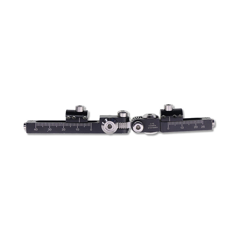
The distal tibia external fixator incorporates sophisticated mechanical components that provide surgeons with unprecedented control over bone position in all three spatial dimensions. This technological advancement represents a fundamental advantage over rigid fixation systems that lock bone fragments in a single position. The adjustable framework features telescoping rods, rotating joints, and calibrated mechanisms that permit gradual correction of angular deformities, rotational malalignment, and limb length discrepancies throughout the healing process. Surgeons can make precise millimeter-level adjustments during follow-up appointments without subjecting patients to additional surgical procedures, reducing healthcare costs and patient discomfort. This capability proves especially valuable when treating complex fracture patterns where initial alignment may be suboptimal due to soft tissue swelling or uncertainty about bone fragment viability. As swelling subsides and treatment progresses, the distal tibia external fixator can be methodically adjusted to optimize bone alignment, ensuring proper union and restoring normal limb mechanics. The ability to dynamically modify fixation parameters based on healing response represents a significant clinical advantage that directly improves patient outcomes and functional recovery.
Superior Soft Tissue Access and Management

Superior Soft Tissue Access and Management

One of the most compelling features of the distal tibia external fixator is its unique ability to provide stable skeletal fixation while maintaining complete access to surrounding soft tissues. Lower leg injuries frequently involve significant skin damage, muscle trauma, and compromised blood supply that require ongoing monitoring and treatment. The external configuration of this fixation system creates an open field around the injury site, allowing healthcare providers to perform wound care, apply advanced dressing systems, and monitor healing progress without disturbing bone stability. This accessibility becomes critically important when treating open fractures where infection prevention depends on thorough wound management and serial debridement procedures. The distal tibia external fixator facilitates collaboration between orthopedic surgeons and plastic surgeons who may need to perform tissue reconstruction, skin grafting, or flap coverage procedures. Unlike internal plates and screws that sit beneath skin and muscle, the external apparatus does not create additional foreign body burden within compromised tissues, reducing infection risk in contaminated wounds. This design philosophy recognizes that successful fracture treatment requires addressing both skeletal and soft tissue components simultaneously, with neither aspect compromising the other.
Rapid Application in Emergency Situations

Rapid Application in Emergency Situations

The distal tibia external fixator excels as a damage control orthopedic solution that can be rapidly deployed in emergency trauma scenarios where immediate stabilization is paramount. Severe lower leg injuries often occur in polytrauma patients who have sustained multiple life-threatening injuries requiring prioritized treatment. The quick application process of the distal tibia external fixator allows trauma surgeons to achieve skeletal stability within minutes, controlling hemorrhage from bone fractures and preventing further soft tissue damage from unstable bone fragments. This rapid stabilization enables patient transport to specialized facilities, permits necessary life-saving procedures to proceed without interference, and provides a stable foundation for subsequent definitive treatment. The technique requires less extensive surgical exposure than internal fixation alternatives, reducing operative time, blood loss, and physiological stress on critically injured patients whose condition may not tolerate lengthy procedures. Once applied, the distal tibia external fixator maintains fracture reduction during the acute injury phase when tissue swelling peaks and wound conditions evolve. Medical teams can defer complex reconstructive procedures until the patient is physiologically optimized and local tissue conditions have improved, following damage control principles that have been proven to reduce complications and mortality in severe trauma cases.
logo