EN
EN

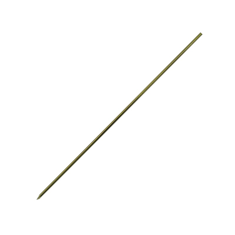
Lisfranc injuries present one of the most challenging fixation problems in foot and ankle surgery, and the choice of implant directly shapes the clinical outcome. Among available fixation tools, the Kirschner wire holds a well-established place in the surgical armamentarium. When a Kirschner wire is used as part of a limited internal fixation strategy, combined with an external fixator frame, surgeons can achieve adequate joint stabilization while minimizing soft-tissue disruption at a site notorious for poor wound healing.
This article examines how effective this combined approach truly is, walking through the biomechanical rationale, surgical technique considerations, and the evidence base that supports or refines the use of a Kirschner wire alongside external fixation for Lisfranc injuries. Understanding when and how a Kirschner wire performs best allows clinicians to make better-informed decisions for this complex injury pattern.
Biomechanical Role of Kirschner Wire in Lisfranc Fixation
How a Kirschner Wire Stabilizes the Tarsometatarsal Complex
The tarsometatarsal joint complex tolerates significant rotational and shear forces during gait, making stable fixation essential after a Lisfranc disruption. A Kirschner wire achieves provisional and sometimes definitive stabilization by traversing the reduced joint surfaces and holding them in anatomical alignment. The smooth shaft of a Kirschner wire allows controlled insertion under fluoroscopic guidance, minimizing articular damage during placement. Because a Kirschner wire is comparatively thin and flexible, it introduces less soft-tissue stripping than bulkier plate-and-screw constructs, which matters greatly in a region where the dorsal skin is already under tension after injury.
When a Kirschner wire is placed across the first, second, or third tarsometatarsal joints in conjunction with an external fixator, the construct distributes load between the two systems. The external fixator neutralizes gross displacement forces while the Kirschner wire maintains precise articular reduction. This load-sharing principle is why the combination often outperforms either technique used alone in high-energy or open Lisfranc injuries.
Kirschner Wire Geometry and Trocar-Point Design
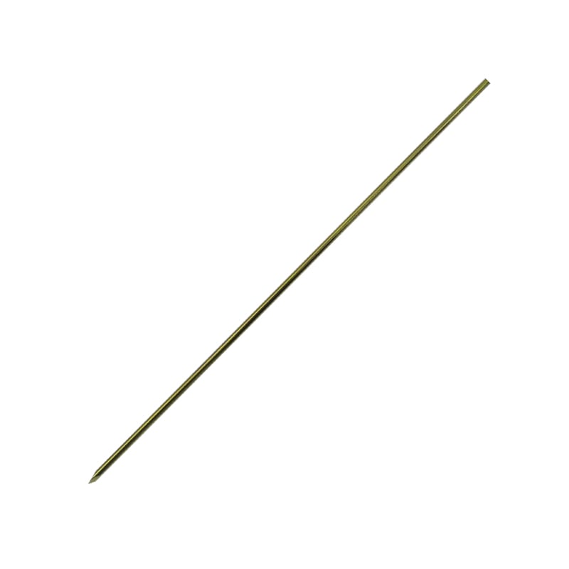
The effectiveness of a Kirschner wire in dense tarsal bone depends significantly on tip geometry. A trocar-point Kirschner wire penetrates cortical bone with less rotational resistance than a diamond-point wire, reducing the risk of wire deflection during insertion. Precise Kirschner wire placement is critical in Lisfranc fixation because even a few millimeters of malalignment can alter tarsometatarsal mechanics. Surgeons therefore prioritize a Kirschner wire design that combines sharpness, torsional rigidity, and consistent diameter tolerance to ensure reproducible placement across multiple joints simultaneously.
Clinical Effectiveness: Evidence and Outcomes
What the Literature Reports on Kirschner Wire Use
Clinical studies evaluating limited internal fixation for Lisfranc injuries consistently demonstrate that a Kirschner wire, when properly positioned and protected by an external frame, can yield satisfactory medium-term outcomes in carefully selected patients. Retrospective series report acceptable rates of anatomical reduction maintenance at 12 weeks, the typical period before a Kirschner wire is removed. Because a Kirschner wire is not designed as a permanent implant, early removal at six to eight weeks is planned in most protocols, which reduces the risk of wire breakage and joint stiffness.
Functional scores in patients treated with a Kirschner wire combined with external fixation are broadly comparable to those treated with primary screw fixation in low-to-moderate energy injuries, although direct comparison remains limited by heterogeneous patient populations. The key advantage of the Kirschner wire approach is its applicability in contaminated wounds, polytrauma settings, and cases with significant soft-tissue compromise where definitive internal fixation must be delayed. In these scenarios, the Kirschner wire serves as a reliable bridge implant that maintains reduction without escalating wound complications.
Limitations and Risk Factors Associated with Kirschner Wire Fixation
No fixation strategy is without limitations, and the Kirschner wire is no exception. Pin-tract infection remains the most reported complication when a Kirschner wire exits through the skin, particularly when combined with an external fixator that also has percutaneous pins. Surgeons mitigate this risk through meticulous pin-site care protocols and by burying the Kirschner wire beneath the skin where the anatomy allows. Loss of reduction is another concern, especially if the Kirschner wire is left in place beyond the recommended duration or if the external fixator is removed prematurely. Careful patient compliance monitoring is therefore integral to a successful Kirschner wire and external fixation protocol.
Surgical Technique Principles for Combined Fixation
Sequencing Kirschner Wire Placement with External Fixation
Optimal outcomes depend on the sequence in which a Kirschner wire and external fixator components are deployed. Most surgeons prefer to achieve gross reduction using the external fixator first, restoring overall foot length and alignment under fluoroscopy. Once the external frame holds the foot in a corrected position, the Kirschner wire is introduced percutaneously across the targeted tarsometatarsal joints to lock in articular reduction. This sequence prevents the Kirschner wire from being stressed by residual deforming forces before the fixator neutralizes them. Each Kirschner wire should be cut close to the skin surface and capped to reduce soft-tissue irritation during the fixation period.
Postoperative Management and Kirschner Wire Removal Timing
After surgery, weight-bearing restrictions are maintained until the Kirschner wire is electively removed, typically at six to eight weeks for most Lisfranc patterns. The removal of a Kirschner wire is straightforward under local or regional anesthesia in an outpatient setting, which is a practical advantage over buried screw hardware that requires a more involved secondary procedure. Following Kirschner wire removal, progressive weight-bearing commences alongside physiotherapy focused on restoring midfoot proprioception and intrinsic muscle strength. The overall rehabilitation timeline when a Kirschner wire is used tends to mirror that of screw-based fixation, though the flexibility of the Kirschner wire protocol allows earlier adjustment if healing is delayed.
FAQ
Is a Kirschner wire strong enough to hold Lisfranc reduction without additional support?
A Kirschner wire alone provides limited rotational stability and is not recommended as standalone fixation for most Lisfranc injuries. The Kirschner wire is most effective when it works in concert with an external fixator or supplementary screw fixation, which together provide the necessary multi-plane stability that the tarsometatarsal complex demands during healing.
How long should a Kirschner wire remain in place after Lisfranc fixation?
Most clinical protocols advocate removing a Kirschner wire at six to eight weeks postoperatively, once early fibrous union stabilizes the reduced joints. Leaving a Kirschner wire beyond ten to twelve weeks increases the risk of wire fatigue fracture and chronic pin-tract infection, both of which complicate the recovery process and may necessitate additional intervention.
Can a Kirschner wire be used for all Lisfranc injury severity grades?
A Kirschner wire is well-suited for low-to-moderate energy Lisfranc injuries, including Myerson types A and B, when combined with appropriate external support. For high-energy, severely comminuted, or purely ligamentous Lisfranc injuries, a Kirschner wire may serve only as a temporary bridge while the surgeon plans definitive screw fixation or primary arthrodesis once soft-tissue conditions allow.