EN

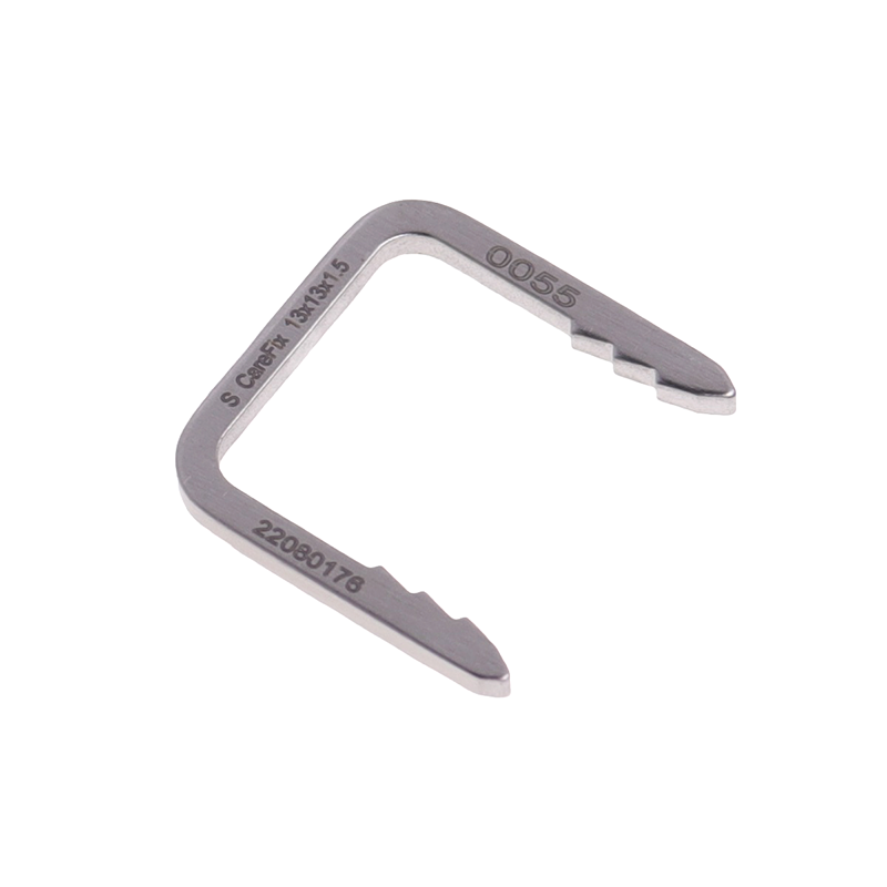
Spinal stabilization requires precise alignment and secure fixation of vertebral segments to promote fusion and prevent harmful motion. Internal fixation systems represent the gold standard for achieving this stability across a wide range of spinal pathologies. Understanding the distinct types of internal fixation devices, their mechanical principles, and their specific clinical indications is essential for orthopedic surgeons, neurosurgeons, and spinal specialists. Each approach offers unique biomechanical advantages and surgical trade-offs that influence patient outcomes, recovery timelines, and long-term fusion success rates.
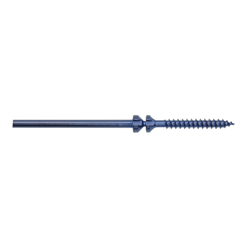
Modern spinal surgery relies on three primary internal fixation strategies: anterior approaches, posterior approaches, and minimally invasive techniques. Each category encompasses specific instrumentation, anatomical exposure requirements, and biomechanical load-sharing characteristics. Selecting the appropriate internal fixation method depends on pathology location, patient anatomy, bone quality, surgeon expertise, and desired fusion architecture. This article explores the essential characteristics, clinical indications, and practical considerations for each major type of internal fixation system in contemporary spine surgery.
Understanding Anterior Spinal Internal Fixation
Mechanism and Biomechanics of Anterior Internal Fixation
Anterior internal fixation systems place instrumentation directly ventral to the vertebral bodies through direct surgical exposure. This approach places load-bearing hardware closer to the center of mass of the spine, creating a more balanced mechanical environment. Anterior internal fixation devices typically include plating systems, cage devices, and hybrid constructs that combine plates with interbody implants. The vertebral bodies themselves carry substantial compressive loads in the cervical and lumbar spine, making anterior internal fixation biomechanically efficient for managing flexion-distraction injuries and degenerative disc disease affecting multiple segments.
Clinical Indications for Anterior Internal Fixation
Anterior internal fixation excels for cervical spondylosis, multilevel degenerative disc disease, anterior vertebral body fractures, and kyphotic deformities requiring reconstruction. Single-level or two-level cervical degenerative conditions represent the most common applications of anterior internal fixation techniques. Anterior internal fixation allows direct decompression of neural elements compressed by disc material or osteophytes while simultaneously restoring disc height and sagittal alignment. Patients with compromised posterior elements, previous posterior surgery, or unfavorable anatomy for dorsal instrumentation often benefit from anterior internal fixation strategies. The direct anterior approach also reduces blood loss and operative time compared to circumferential fusion requiring both anterior and posterior exposure.
Posterior Spinal Internal Fixation Systems
Screw-Rod Construct Design and Load Distribution
Posterior internal fixation systems dominate modern spinal surgery, utilizing pedicle screws connected by longitudinal rods to create rigid three-dimensional constructs. This architecture provides excellent resistance to flexion, extension, and shear forces while maintaining segmental motion control across injured or degenerative segments. Posterior internal fixation constructs can span multiple vertebral levels and accommodate different vertebral anatomy from cervical through sacral regions. The internal fixation principles of pedicle screw-based systems create strong cantilever constructs that resist both anterior and posterior loads, making them suitable for complex traumatic injuries and severe degenerative pathologies.
Indications for Posterior Internal Fixation
Posterior internal fixation serves as the primary fixation method for traumatic fracture-dislocations, burst fractures with retropulsion, spondylolisthesis, and severe degenerative spondylosis with instability. Multi-level lumbar degenerative disc disease with stenosis and listlessness frequently requires posterior internal fixation for adequate stabilization. Posterior internal fixation systems excel in high-grade spondylolisthesis where multiple segments require segmental reduction and stabilization. Adolescent idiopathic scoliosis treatment relies heavily on posterior internal fixation technology to achieve and maintain correction. Tumors, infections, and inflammatory conditions affecting posterior elements or requiring sacrifice of stabilizing structures often mandate posterior internal fixation approaches. The posterior anatomy generally provides stronger bone purchase for pedicle screw engagement compared to anterior vertebral body bone quality in elderly or osteoporotic patients.
Minimally Invasive Internal Fixation Approaches
Percutaneous and Tubular Techniques for Internal Fixation
Minimally invasive internal fixation techniques utilize smaller incisions, percutaneous screw placement, and tubular retractor systems to reduce soft tissue trauma while maintaining fixation quality comparable to open approaches. Minimally invasive internal fixation methods preserve posterior musculature, reduce blood loss, and accelerate rehabilitation timelines. Percutaneous pedicle screw technology allows three-dimensional trajectory planning and fluoroscopic guidance to ensure accurate screw positioning within constrained anatomical windows. Minimally invasive internal fixation systems incorporate specialized instrumentation, including lateral mass screws, translaminar screws, and percutaneous rod placement techniques. The learning curve for minimally invasive internal fixation procedures exceeds that of open techniques, requiring dedicated training and specialized imaging intraoperatively.
Indications and Patient Selection for Minimally Invasive Internal Fixation
Minimally invasive internal fixation suits patients with focal pathology amenable to limited fusion, elderly individuals with significant comorbidities, and those desiring faster recovery timelines. Single-level and two-level degenerative conditions represent ideal candidates for minimally invasive internal fixation techniques. Minimally invasive internal fixation approaches benefit patients with obesity, revision surgery history, or extensive posterior scar tissue where open exposure proves technically challenging. Acute traumatic fractures with severe soft tissue injury often justify minimally invasive internal fixation selection to minimize additional iatrogenic damage. Minimally invasive internal fixation techniques demonstrate higher accuracy and safety when limited to single or dual-segment constructs, as wider exposure challenges increase technical difficulty.
FAQ
What determines whether anterior or posterior internal fixation is optimal for a specific patient?
Pathology location, deformity pattern, bone quality, and patient comorbidities guide internal fixation selection. Anterior internal fixation suits ventral pathology and kyphotic deformities, while posterior internal fixation excels for dorsal element involvement, spondylolisthesis, and multi-level instability. Patient anatomy, previous surgical history, and surgeon expertise also influence internal fixation approach choice significantly.
Can minimally invasive internal fixation achieve equivalent fusion rates compared to open internal fixation procedures?
Yes, minimally invasive internal fixation demonstrates comparable fusion rates to open internal fixation when applied to appropriate single or dual-segment pathology. Long-term studies show equivalent bone union, reduced infection rates, and fewer complications with minimally invasive internal fixation in suitable candidates. However, internal fixation outcomes depend on patient selection, technical accuracy, and adequate bony contact rather than surgical approach alone.
How does bone quality influence internal fixation system selection and implant sizing?
Osteoporotic bone limits pull-out strength for internal fixation screws, necessitating wider implant diameters or augmentation techniques like cement to enhance purchase. Internal fixation systems in poor-quality bone may require longer construct lengths to distribute loads across additional segments. Patients with compromised bone quality often benefit from internal fixation techniques prioritizing larger surface area contact and avoiding sole reliance on screw-based constructs.
Table of Contents
- Understanding Anterior Spinal Internal Fixation
- Posterior Spinal Internal Fixation Systems
- Minimally Invasive Internal Fixation Approaches
-
FAQ
- What determines whether anterior or posterior internal fixation is optimal for a specific patient?
- Can minimally invasive internal fixation achieve equivalent fusion rates compared to open internal fixation procedures?
- How does bone quality influence internal fixation system selection and implant sizing?