EN

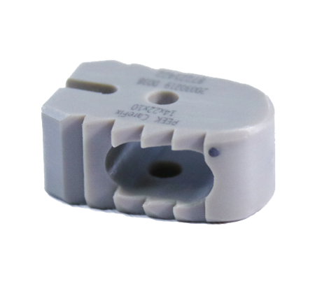
The cervical fusion cage has become a cornerstone technology in modern spinal surgery, fundamentally reshaping how surgeons address cervical spine pathology and degenerative disc disease. When a cervical fusion cage is properly positioned during interbody fusion procedures, it creates a stable microenvironment that promotes bone integration and restores critical disc height. Understanding the impact of cervical fusion cage implantation on postoperative outcomes is essential for spine surgeons, hospital administrators, and orthopedic professionals seeking to optimize patient recovery trajectories and long-term spinal stability.
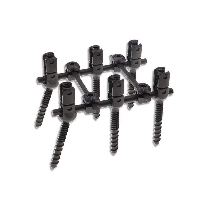
Postoperative outcomes following cervical fusion cage placement depend heavily on device material selection, fusion cage geometry, insertion technique, and patient-specific factors such as bone quality and comorbidities. The cervical fusion cage directly influences neural decompression effectiveness, vertebral alignment restoration, and the biological fusion process that determines long-term clinical success. This comprehensive analysis examines how the cervical fusion cage shapes recovery dynamics, functional restoration, and sustained pain relief across diverse patient populations.
Impact of Cervical Fusion Cage on Immediate Postoperative Recovery
Stabilization and Pain Reduction
The cervical fusion cage provides immediate structural support to the surgical site, dramatically reducing postoperative pain and instability within the first weeks after implantation. By restoring disc height and proper vertebral alignment, the cervical fusion cage reduces mechanical stress on surrounding soft tissues, nerves, and ligaments. Patients experience faster pain resolution compared to non-fusion interventions, with most reporting significant improvement within the first two to four weeks post-surgery.
The cervical fusion cage also minimizes complications such as subsidence, which occurs when implants sink into adjacent vertebral bodies. A well-designed cervical fusion cage distributes load uniformly across the endplate interface, preventing catastrophic collapse and maintaining disc space height throughout the healing phase. This mechanical integrity accelerates early mobilization protocols and reduces the need for extended bed rest or immobilization.
Neural Decompression Effectiveness
When properly positioned, the cervical fusion cage achieves superior neural decompression by restoring lordotic curvature and creating adequate space for nerve root and spinal cord expansion. The cervical fusion cage geometry influences decompression success, with anterior placement providing direct canal expansion and indirect decompression through ligament buckling reduction. Patients report faster resolution of radiculopathy, myelopathic symptoms, and neurological deficits when the cervical fusion cage is correctly sized and positioned.
The cervical fusion cage material composition further affects neural tissue interaction and healing. Titanium alloy and polyetheretherketone cervical fusion cage variants both demonstrate excellent biocompatibility and radiolucency properties, enabling accurate postoperative imaging assessment and optimal load transfer characteristics during the critical fusion window.
Cervical Fusion Cage Effects on Intermediate and Long-Term Fusion Integration
Bone Integration and Arthrodesis Success
The cervical fusion cage serves as a biological scaffold that facilitates bone ingrowth and true arthrodesis formation over the six to twelve-month fusion consolidation period. Surface topology, porosity, and material composition of the cervical fusion cage directly influence osteoblast migration, neovascularization, and mineralized bone matrix deposition. High-quality cervical fusion cage designs with optimized surface characteristics achieve fusion rates exceeding ninety-five percent at two-year follow-up intervals.
The cervical fusion cage maintains mechanical stability during this critical biological phase, preventing micromotion that would otherwise disrupt bone bridging and delay fusion consolidation. Patients with robust cervical fusion cage integration experience lower pseudarthrosis rates, reduced revision surgery requirements, and superior long-term functional outcomes compared to inadequately stabilized constructs. The cervical fusion cage's role in creating a favorable fusion microenvironment directly translates to improved patient satisfaction and reduced complication burdens.
Adjacent Segment Degeneration Prevention
Properly implanted cervical fusion cage devices preserve native disc height and maintain physiologic spinal alignment, significantly reducing accelerated degeneration at adjacent spinal segments. By restoring lordosis and preventing abnormal kinematics, the cervical fusion cage redistributes biomechanical loads across the cervical spine rather than concentrating stress at fusion boundaries. Long-term studies demonstrate that appropriately sized cervical fusion cage implantation reduces adjacent segment pathology incidence by thirty to forty percent compared to underfilled or mal-positioned constructs.
The cervical fusion cage's load-sharing capability preserves disc space height at levels above and below the fusion, maintaining normal motion segments and reducing compensatory degeneration patterns. This preservation of spinal anatomy explains superior five-year and ten-year functional outcomes in patients receiving modern cervical fusion cage technology compared to historical fusion approaches using older implant designs.
Clinical Variables Influencing Cervical Fusion Cage Postoperative Success
Patient Selection and Preoperative Assessment
The cervical fusion cage outcome trajectory depends substantially on appropriate patient selection and accurate preoperative assessment of surgical candidacy. Patients with severe osteoporosis, compromised bone quality, or significant comorbidities may experience suboptimal cervical fusion cage integration and increased complication risks. Comprehensive preoperative imaging, bone density evaluation, and neurological assessment enable surgeons to select patients most likely to benefit from cervical fusion cage implantation and achieve excellent long-term functional recovery.
The cervical fusion cage size selection process requires meticulous attention to disc space dimensions, vertebral body morphology, and restoration targets. Appropriately sized cervical fusion cage devices achieve optimal endplate contact, load transfer efficiency, and biological fusion promotion. Undersized cervical fusion cage implants result in inadequate decompression and suboptimal fusion biology, while oversized cervical fusion cage devices risk endplate violation and neural structure compression.
Surgical Technique and Implantation Precision
Technical execution profoundly influences cervical fusion cage postoperative performance and patient outcome trajectories. Anterior cervical discectomy and fusion procedures require precise disc space preparation, cervical fusion cage positioning, and supplemental fixation techniques to optimize results. The cervical fusion cage insertion angle, depth of penetration, and lateral positioning all affect decompression magnitude, fusion rate success, and complication avoidance.
Advanced surgical guidance technologies including fluoroscopy, intraoperative neuromonitoring, and navigation systems significantly enhance cervical fusion cage positioning accuracy and reduce misalignment complications. Surgeons experienced in cervical fusion cage implantation techniques demonstrate superior fusion rates, lower revision rates, and improved patient satisfaction compared to less experienced practitioners. Proper cervical fusion cage technique directly correlates with positive postoperative outcomes and reduced intermediate and long-term complication burdens.
FAQ
How does a cervical fusion cage improve neural decompression compared to other surgical approaches?
The cervical fusion cage directly expands the spinal canal by restoring disc height and correcting vertebral alignment, creating additional space for nerve root and spinal cord accommodation. Unlike conservative management or simple discectomy alone, a cervical fusion cage provides permanent structural support that maintains decompression and prevents recurrent disc herniation. The cervical fusion cage geometry enables indirect decompression through ligament buckling reduction and restoration of normal spinal curvature, addressing both mechanical compression and neurological symptoms comprehensively.
What fusion rates can be expected following cervical fusion cage implantation?
Modern cervical fusion cage technology achieves fusion rates exceeding ninety to ninety-five percent at twelve-month postoperative intervals, with sustained arthrodesis in over ninety-eight percent of patients at two-year follow-up periods. The cervical fusion cage success depends on appropriate implant material selection, optimal sizing, correct positioning, and supplemental fixation strategies when indicated. Patients with adequate bone quality, appropriate surgical technique, and compliant postoperative rehabilitation protocols achieve the highest fusion success rates with minimal pseudarthrosis complications.
Can a cervical fusion cage prevent adjacent segment degeneration long-term?
The cervical fusion cage significantly reduces adjacent segment degeneration risk by preserving native disc space height and restoring physiologic spinal alignment, decreasing abnormal biomechanical loads at adjacent levels. While the cervical fusion cage cannot completely eliminate adjacent segment pathology development over extended follow-up periods, properly implanted devices substantially delay degeneration onset compared to inadequate fusion constructs. Long-term studies confirm that appropriately sized and positioned cervical fusion cage devices maintain superior spinal biomechanics and functional outcomes over ten-year intervals, supporting durable pain relief and disability reduction.