
The evolution of pediatric orthopedic surgery has witnessed remarkable advancements in recent decades, with the telescopic intramedullary nail emerging as a groundbreaking solution for treating femoral shaft fractures in growing children. This innovative medical device addresses one of the most challenging aspects of pediatric fracture management: accommodating continued bone growth while maintaining optimal fracture stabilization. Traditional rigid intramedullary nails often required secondary surgical procedures to prevent complications as children grew, but the telescopic intramedullary nail has revolutionized this approach by providing dynamic length adjustment that synchronizes seamlessly with natural bone development.
Understanding the Mechanics of Telescopic Technology
Core Design Principles
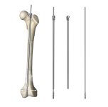
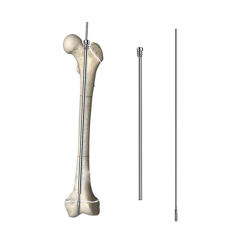
The telescopic intramedullary nail operates on sophisticated engineering principles that allow controlled expansion within the medullary canal of long bones. The device consists of two primary components: an outer sleeve and an inner rod that can slide within the sleeve mechanism. This telescoping action is facilitated by precision-engineered internal mechanisms that respond to natural physiological forces generated during bone growth and remodeling processes.
The outer diameter of the telescopic intramedullary nail is carefully calculated to provide optimal contact with the endosteal surface while allowing sufficient space for the expansion mechanism. The internal sliding components are manufactured from biocompatible materials that resist corrosion and maintain smooth operation throughout the device's functional lifespan. Advanced surface treatments ensure minimal friction between moving parts, enabling the telescopic intramedullary nail to extend gradually as bone growth occurs.
Biomechanical Adaptation Mechanisms
The synchronization between the telescopic intramedullary nail and bone growth relies on biomechanical feedback loops that naturally occur during skeletal development. As the child grows, longitudinal forces generated by muscle contractions, weight-bearing activities, and normal physiological stress create controlled tension within the nail system. These forces trigger the telescoping mechanism, allowing gradual extension that matches the rate of bone elongation.
Research demonstrates that the telescopic intramedullary nail responds proportionally to growth stimuli, with extension rates typically ranging from 0.5 to 2 millimeters per month depending on the child's age and growth velocity. This adaptive response ensures that the nail maintains proper positioning within the medullary canal while providing continuous stabilization throughout the healing and growth phases. The mechanism's sensitivity to physiological forces prevents premature or excessive extension while ensuring adequate responsiveness to legitimate growth demands.
Clinical Applications and Patient Selection
Optimal Candidate Characteristics
Selection of appropriate candidates for telescopic intramedullary nail implantation requires careful consideration of multiple factors including age, growth potential, fracture pattern, and overall health status. Children between ages 6 and 14 typically represent ideal candidates, as this age range corresponds to significant remaining growth potential while ensuring sufficient bone diameter to accommodate the device. The telescopic intramedullary nail performs optimally in patients with at least 2-3 years of expected growth remaining.
Fracture characteristics also influence candidacy, with transverse and short oblique femoral shaft fractures responding most favorably to telescopic intramedullary nail fixation. Complex fracture patterns, significant comminution, or associated injuries may require alternative treatment approaches. Bone quality assessment is crucial, as adequate cortical thickness and bone density ensure proper nail engagement and stability throughout the growth period.
Surgical Technique Considerations
Implantation of the telescopic intramedullary nail requires specialized surgical techniques that differ significantly from traditional rigid nail procedures. The entry point selection must account for future growth patterns, typically utilizing a trochanteric entry to avoid damage to the femoral head blood supply. Reaming procedures are modified to accommodate the larger diameter of the telescopic intramedullary nail while preserving endosteal blood supply essential for bone healing.
Intraoperative positioning of the telescopic intramedullary nail requires precise calculation of initial length settings to ensure adequate expansion capacity throughout the expected growth period. Surgeons must consider the patient's growth velocity, remaining growth potential, and desired final nail position when determining initial telescoping settings. Advanced imaging techniques guide optimal placement and confirm proper mechanical alignment before wound closure.
Growth Synchronization Mechanisms
Physiological Growth Monitoring
The telescopic intramedullary nail incorporates sophisticated monitoring capabilities that allow real-time assessment of growth progression and mechanical performance. Radiopaque markers within the device enable radiographic measurement of extension distance during routine follow-up examinations. These measurements provide quantitative data regarding growth rates and help clinicians verify proper synchronization between bone development and nail expansion.
Growth velocity calculations derived from serial radiographic measurements help predict future expansion requirements and identify potential complications before they become clinically significant. The telescopic intramedullary nail's response to growth stimuli can be tracked and compared to normal growth curves, ensuring that the device maintains optimal function throughout the treatment period. Any deviation from expected extension patterns triggers enhanced monitoring protocols and potential intervention planning.
Adaptive Response Mechanisms
The telescopic intramedullary nail demonstrates remarkable adaptive capabilities that allow automatic adjustment to varying growth rates and mechanical demands. During periods of rapid growth, typically occurring during pubertal growth spurts, the device increases its extension rate to maintain proper positioning within the elongating bone. Conversely, during slower growth phases, the telescopic intramedullary nail reduces its extension rate to prevent over-lengthening.
This adaptive response is mediated through mechanical feedback systems that detect changes in axial loading patterns and bone remodeling activity. Increased osteoblastic activity associated with rapid growth generates enhanced mechanical stimuli that trigger more aggressive nail extension. The telescopic intramedullary nail's ability to modulate its response ensures consistent synchronization with natural bone development patterns throughout diverse growth phases.
Advantages Over Traditional Treatment Methods
Elimination of Secondary Procedures
Perhaps the most significant advantage of the telescopic intramedullary nail lies in its ability to eliminate the need for secondary surgical procedures traditionally required with rigid nail systems. Conventional intramedullary nails often necessitate removal and replacement as children grow, exposing patients to additional surgical risks, anesthesia complications, and prolonged recovery periods. The telescopic intramedullary nail's self-adjusting mechanism obviates these concerns by providing continuous adaptation throughout the growth period.
This elimination of secondary procedures translates to substantial reductions in healthcare costs, patient morbidity, and family disruption. Parents and children benefit from the psychological relief of knowing that additional surgeries are typically unnecessary, reducing anxiety and improving overall treatment satisfaction. The telescopic intramedullary nail's longevity also minimizes the risk of complications associated with multiple surgical interventions, including infection, blood loss, and anesthetic exposure.
Enhanced Functional Outcomes
Clinical studies demonstrate superior functional outcomes in patients treated with telescopic intramedullary nail systems compared to traditional rigid nail approaches. The device's ability to maintain optimal mechanical alignment throughout growth periods results in improved limb length equality, reduced angular deformities, and enhanced overall function. Patients experience faster return to normal activities and reduced long-term disability rates.
The telescopic intramedullary nail's dynamic properties also contribute to improved bone remodeling and strength development. By maintaining physiological loading patterns throughout healing and growth, the device promotes normal bone architecture development and mineral density optimization. This results in stronger, more resilient bone structures that better resist future injury and maintain function throughout the patient's lifetime.
Long-term Performance and Durability
Material Science Innovations
The telescopic intramedullary nail incorporates advanced materials science innovations that ensure exceptional durability and biocompatibility throughout extended implantation periods. Titanium alloy construction provides optimal strength-to-weight ratios while maintaining excellent corrosion resistance in the physiological environment. Specialized surface treatments minimize wear debris generation and reduce the risk of adverse tissue reactions.
Advanced manufacturing techniques ensure precise tolerances between moving components, enabling smooth telescoping action throughout the device's functional lifespan. Quality control procedures verify that each telescopic intramedullary nail meets stringent performance specifications for extension force, fatigue resistance, and dimensional stability. These manufacturing standards ensure consistent clinical performance across all device units.
Longevity and Replacement Considerations
Long-term follow-up studies indicate that the telescopic intramedullary nail can function effectively for periods of 5-10 years or more, often encompassing the entire remaining growth period in pediatric patients. The device's robust construction and reliable telescoping mechanisms rarely require premature replacement due to mechanical failure. Most telescopic intramedullary nail removals occur after growth completion rather than due to device malfunction.
When removal becomes necessary, typically after skeletal maturity, the telescopic intramedullary nail can be extracted using standard surgical techniques. The extended implantation period allows complete bone healing and remodeling, often resulting in near-normal bone architecture at the time of removal. Patients who undergo nail removal after growth completion typically experience excellent long-term outcomes with minimal functional limitations.
Future Developments and Innovations
Smart Technology Integration
The next generation of telescopic intramedullary nail systems is expected to incorporate smart technology features that further enhance monitoring capabilities and treatment outcomes. Integrated sensors may provide real-time data regarding mechanical loading, extension rates, and bone healing progress. These technological advances will enable more precise treatment optimization and earlier detection of potential complications.
Wireless communication capabilities may allow remote monitoring of telescopic intramedullary nail performance, reducing the frequency of clinical visits while maintaining comprehensive oversight of treatment progress. Advanced algorithms could analyze sensor data to predict optimal extension patterns and alert clinicians to any deviations from expected performance parameters. These innovations represent the future of personalized orthopedic care in pediatric populations.
Expanded Clinical Applications
Research continues to explore expanded applications for telescopic intramedullary nail technology beyond femoral shaft fractures. Potential applications include tibial fractures, humerus injuries, and congenital limb length discrepancies. The underlying telescoping principles may also be adapted for other orthopedic devices, including external fixators and joint replacement components for growing children.
International collaborative studies are investigating optimal patient selection criteria, refined surgical techniques, and enhanced device designs that could further improve outcomes. The telescopic intramedullary nail continues to evolve as our understanding of pediatric bone biology and biomechanics advances, promising even better treatment options for future generations of young patients.
FAQ
How long does a telescopic intramedullary nail remain functional in a growing child
A telescopic intramedullary nail typically remains functional throughout the entire remaining growth period in pediatric patients, which can range from 2 to 8 years depending on the child's age at implantation. The device is designed to accommodate the total expected growth of the femur, with most nails providing 4-6 centimeters of extension capability. Clinical studies show that over 95% of telescopic intramedullary nail implants function properly until growth completion without requiring replacement or revision surgery.
What are the main differences between telescopic and traditional intramedullary nails
The primary difference lies in the telescopic intramedullary nail's ability to extend automatically as the bone grows, whereas traditional rigid nails maintain a fixed length. Traditional nails often require removal and replacement with longer nails as children grow, typically necessitating 1-3 additional surgeries. Telescopic nails eliminate this need through their self-adjusting mechanism that responds to natural growth forces. Additionally, telescopic nails are specifically designed for pediatric patients, while traditional nails are primarily used in adults with completed skeletal growth.
Are there any activity restrictions for children with telescopic intramedullary nails
Children with telescopic intramedullary nails can typically return to most normal activities within 2-3 months after surgery, including running, cycling, and recreational sports. However, high-impact activities such as contact sports, gymnastics, or activities with high fall risk may require longer restriction periods or permanent limitation depending on individual circumstances. The telescopic mechanism actually benefits from normal loading and activity, as physiological forces help drive the extension process. Most children can participate in school physical education and organized sports with appropriate modifications and protective equipment.
How do surgeons monitor the extension progress of telescopic intramedullary nails
Surgeons monitor telescopic intramedullary nail extension through regular radiographic examinations, typically performed every 3-6 months during the active growth period. The nail contains radiopaque markers that allow precise measurement of extension distance on X-rays. These measurements are compared to the child's overall growth velocity and expected bone length to ensure proper synchronization. Advanced imaging techniques may also be used to assess bone healing progress and verify optimal nail positioning within the medullary canal throughout the treatment period.
Table of Contents
- Understanding the Mechanics of Telescopic Technology
- Clinical Applications and Patient Selection
- Growth Synchronization Mechanisms
- Advantages Over Traditional Treatment Methods
- Long-term Performance and Durability
- Future Developments and Innovations
-
FAQ
- How long does a telescopic intramedullary nail remain functional in a growing child
- What are the main differences between telescopic and traditional intramedullary nails
- Are there any activity restrictions for children with telescopic intramedullary nails
- How do surgeons monitor the extension progress of telescopic intramedullary nails
