EN

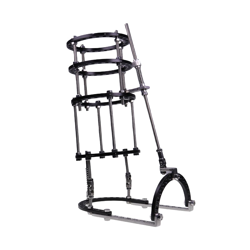
The Ilizarov brace represents one of the most versatile and effective approaches in modern orthopedic surgery, offering clinicians and patients a proven method for addressing complex skeletal deformities, fracture complications, and limb reconstruction challenges. Ring external fixation, the core principle underlying Ilizarov technology, has revolutionized how orthopedic teams manage cases that were previously considered difficult or impossible to treat with traditional methods. This comprehensive guide explores the multi-scenario treatment advantages of ring external fixation and why the Ilizarov system remains the gold standard for precision limb correction across diverse clinical presentations.

What sets ring external fixation apart from other fixation methods is its exceptional biomechanical stability combined with the ability to apply gradual, three-dimensional correction. Unlike plate-and-screw systems or traditional linear fixators, ring external fixation distributes loads across a wider surface area through multiple attachment points, creating a more forgiving construct that accommodates soft tissue healing and patient mobility during the treatment phase. This adaptability makes ring external fixation particularly valuable in trauma settings, where unpredictable complications often emerge weeks or months after initial injury.
Ring External Fixation for Trauma and Fracture Management
Acute Fracture Stabilization and Complication Resolution
Traumatic fractures, especially those involving the tibia, femur, and ankle, frequently present with comminution, soft tissue damage, and vascular compromise that make traditional fixation risky or ineffective. Ring external fixation provides immediate stability without requiring extensive soft tissue stripping, which is critical when swelling and contamination are present. Surgeons can apply ring external fixation in the acute phase, stabilize the fracture, and then transition to definitive treatment once the soft tissue envelope has recovered and the patient is more stable.
When fracture complications such as malunion, nonunion, or infection develop, ring external fixation becomes an invaluable salvage option. The adjustability of ring external fixation allows surgeons to correct both the fracture site and any emerging malalignment without reopening the original injury. This staged approach reduces infection risk and improves patient outcomes compared to revision open reduction and internal fixation, especially in contaminated or infected bone.
Periosteal Preservation and Biological Healing
One of the most significant advantages of ring external fixation is that it preserves the periosteum and avoids disrupting the fracture hematoma. By maintaining the biological environment around the fracture, ring external fixation promotes faster bone healing and reduces the risk of delayed union or nonunion. This biological advantage becomes increasingly important in older patients, in those with compromised healing capacity, or in polytrauma cases where multiple injuries tax the body's ability to heal.
Ring External Fixation for Deformity Correction and Limb Reconstruction
Gradual Distraction and Three-Dimensional Correction
Congenital limb deformities, acquired angulation, length discrepancies, and rotational malalignment all benefit from the precision control offered by ring external fixation. The Ilizarov principle of gradual distraction, typically advanced at 1 millimeter per day, allows the bone, soft tissues, muscles, and nerves to adapt together without ischemic damage or sudden neurovascular compromise. Ring external fixation can simultaneously correct frontal plane angulation, sagittal plane angulation, rotation, and length discrepancy through a single construct, something few other systems can achieve with the same level of control.
In pediatric cases where growth potential and long-term function are paramount, ring external fixation offers a low-morbidity approach to managing developmental deformities. The ability to make micro-adjustments throughout the treatment period means corrections can be fine-tuned based on real-time radiographic feedback, reducing the likelihood of overcorrection or undercorrection. This precision translates into better functional outcomes and lower reoperation rates compared to one-stage corrective osteotomies.
Segmental Bone Loss and Reconstruction
When patients present with significant bone loss from trauma, infection, or tumor resection, ring external fixation enables bone transport and segmental reconstruction that would otherwise require complex vascularized bone grafts or free flap surgery. The ring external fixation framework can house a corticotomy site that is gradually distracted to generate new bone, which then fills the defect zone. This distraction osteogenesis technique using ring external fixation has enabled limb salvage and restoration of function in cases previously destined for amputation.
Ring External Fixation for Infection Control and Soft Tissue Management
Access and Wound Care During Treatment
Infected nonunions, osteomyelitis, and complex wounds benefit enormously from ring external fixation because the fixation construct remains external and does not compete for soft tissue space or interfere with wound care. Nurses and clinicians can easily access the affected area, perform dressing changes, apply topical medications, and monitor healing without removing or repositioning the fixation device. This accessibility is particularly important in contaminated or immunocompromised scenarios where frequent wound inspection and intervention are medically necessary.
The minimal soft tissue violation associated with ring external fixation reduces the bacterial bioburden compared to plates, screws, or intramedullary devices, all of which can serve as foreign bodies that harbor infection. In infected bone, ring external fixation's biological approach to healing, combined with the ability to manage the defect surgically, creates an environment where antibiotics are more effective and recurrence rates are lower.
Vascular and Neurological Safety
In limbs with compromised vascularity, extensive scarring, or previous injury, ring external fixation avoids the need for plate positioning or intramedullary reaming that might compromise blood supply. The construct's configuration can be tailored to avoid critical vascular channels and major nerve trunks, which is especially important in the leg and ankle where the posterior tibial vessels and tibial nerve are at risk with certain implant positions. This spatial freedom makes ring external fixation the safer choice in high-risk vascular cases.
FAQ
What makes ring external fixation superior to plate fixation for complex fractures?
Ring external fixation offers superior biological healing because it preserves the periosteum and fracture hematoma, whereas plate fixation requires surgical exposure and soft tissue stripping. Ring external fixation is also more forgiving of malalignment, as corrections can be made gradually throughout treatment without reoperation. In contaminated or infected bone, ring external fixation provides better access for wound care and reduces the risk of implant-related infection compared to internal hardware.
How long does ring external fixation treatment typically take?
Treatment duration depends on the nature and severity of the condition. Simple fracture stabilization may require 6 to 12 weeks, while complex deformity corrections using distraction osteogenesis can take 3 to 6 months or longer. Factors such as patient age, bone healing capacity, infection status, and the extent of correction needed all influence the timeline. Your surgeon will establish a detailed treatment plan that outlines the expected duration for your specific case.
Can ring external fixation be used in pediatric patients?
Yes, ring external fixation is particularly valuable in children because it preserves growth potential and allows gradual correction that accommodates rapid bone remodeling in young patients. The reduced soft tissue trauma and non-invasive nature of ring external fixation make it an excellent first-line choice for many pediatric deformities and fracture complications. The key is careful pin placement away from growth plates and close follow-up to ensure corrections are proceeding appropriately.