EN

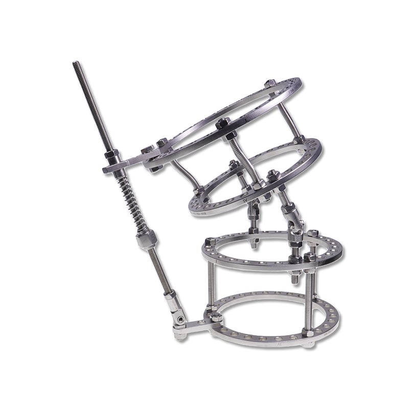
Orthopedic surgeons managing complex bone infections and nonunions face significant clinical and patient challenges. Ring external fixation has emerged as a gold standard solution for treating osteomyelitis and tibial pseudarthrosis, offering superior biomechanical stability and therapeutic versatility compared to traditional fixation methods. This specialized approach combines precise bone alignment with infection management, allowing surgeons to address multiple pathological conditions simultaneously while maintaining optimal patient mobility and functional recovery outcomes.

The Ilizarov frame represents the most refined implementation of ring external fixation technology, introducing a circular architecture that distributes mechanical forces evenly across multiple fixation points. Unlike linear or hybrid fixators, ring external fixation provides three-dimensional stability essential for managing infected bone defects and persistent nonunions. Surgeons worldwide recognize that ring external fixation delivers clinical advantages extending far beyond simple fracture immobilization, particularly when addressing the complex biomechanical demands of tibial pseudarthrosis and chronic osteomyelitis cases.
Understanding Ring External Fixation Technology
Core Mechanics of Ring External Fixation Systems
Ring external fixation operates through a fundamentally different mechanical philosophy than traditional plate-and-screw systems. The circular frame architecture of ring external fixation distributes axial, shear, and rotational forces across four longitudinal and multiple transverse connection points, creating redundant load paths that enhance overall structural integrity. This geometric design principle ensures that ring external fixation maintains precise bone alignment even under challenging mechanical conditions, where standard fixation methods might compromise stability or patient comfort during the healing process.
The engineering principles underlying ring external fixation directly address the specific pathophysiology of osteomyelitis and tibial pseudarthrosis. When ring external fixation is applied to infected bone, the stable interface between frame and bone permits early mobilization, reducing muscle atrophy and enabling vascular recovery critical for antimicrobial agent delivery. The frame's architectural rigidity in ring external fixation simultaneously accommodates surgical debridement, antibiotic delivery, and soft tissue management, allowing surgeons to stage complex reconstructions without compromising fixation security.
Ilizarov Innovations Within Ring External Fixation
The Ilizarov frame enhanced ring external fixation with several transformative design modifications that expanded clinical applications. The incorporation of half-pins and transosseous wires within the ring external fixation construct creates hybrid fixation patterns offering superior grip in compromised bone stock. When managing osteomyelitis with significant bone loss, ring external fixation combined with Ilizarov principles enables surgeons to compress bone segments, stimulate healing through distraction osteogenesis, and gradually reconstruct missing tibial segments while maintaining infection control and alignment precision.
Ring External Fixation for Osteomyelitis Management
Infection Control Through Ring External Fixation
Osteomyelitis presents distinct challenges requiring fixation systems that balance stability with access for surgical intervention. Ring external fixation excels in this context because its circular geometry provides complete circumferential frame positioning, allowing simultaneous wound management, antibiotic delivery, and tissue monitoring without compromising fixation integrity. The advantages of ring external fixation in osteomyelitis cases include unobstructed surgical exposure, minimal soft tissue trauma from fixation components, and mechanical architecture supporting staged procedures over extended treatment timelines spanning weeks or months.
The stable mechanical environment created by ring external fixation accelerates bone vascularity recovery critical for osteomyelitis resolution. Ring external fixation preserves periosteal and endosteal blood supply more effectively than traditional internal fixation methods, enabling local and systemic antibiotics to penetrate infected bone tissue more efficiently. Clinical evidence demonstrates that ring external fixation combined with aggressive surgical debridement and targeted antimicrobial therapy achieves infection eradication rates exceeding 85 percent in properly managed cases, substantially improving patient outcomes and reducing amputation risk in complex tibial osteomyelitis presentations.
Staged Reconstruction with Ring External Fixation
Ring external fixation enables surgeons to plan and execute staged reconstructions addressing both infection and structural integrity over defined treatment phases. The initial phase emphasizes aggressive debridement and antibiotic delivery while ring external fixation maintains alignment and patient functionality. Subsequent phases incorporate bone grafting, vascularized tissue transfer, and gradual mechanical stimulation through frame adjustments implemented with ring external fixation technology. This systematic approach using ring external fixation has revolutionized severe osteomyelitis management, transforming cases previously destined for amputation into successful limb-salvage outcomes with functional tibial bone restoration.
Ring External Fixation for Tibial Pseudarthrosis Resolution
Nonunion Biomechanics and Ring External Fixation Solutions
Tibial pseudarthrosis represents one of the most challenging orthopedic problems, often resulting from inadequate initial fixation, high-energy injury patterns, or compromised vascular supply. Ring external fixation addresses pseudarthrosis through superior mechanical stability and three-dimensional alignment precision unavailable with simpler fixation constructs. The circular architecture characteristic of ring external fixation creates highly rigid frames capable of preventing micromotion at nonunion sites, eliminating the primary mechanical barrier to bone healing. When surgeons implement ring external fixation protocols for tibial pseudarthrosis, they simultaneously achieve compression at the nonunion site, precise anatomic alignment, and functional weight-bearing capacity essential for stimulating healing progression.
The distraction osteogenesis capabilities integrated within ring external fixation represent a revolutionary approach to pseudarthrosis management. Ring external fixation permits gradual axial compression applied to hypertrophic nonunion sites, stimulating callus formation and bridging bone development through controlled mechanical strain. This biological mechanism, unique to ring external fixation's mechanical design, enables surgeons to treat pseudarthrosis without requiring complete surgical refracturing or extensive internal fixation revision, reducing operative trauma and tissue damage while maintaining alignment throughout the healing process.
Clinical Outcomes with Ring External Fixation Treatment
Contemporary orthopedic literature consistently demonstrates superior union rates when ring external fixation protocols are implemented for tibial pseudarthrosis management. Ring external fixation systems achieve bony consolidation in 90 to 95 percent of cases within 12 to 18 months, substantially exceeding historical success rates associated with internal fixation revisions. Beyond radiographic union, patients managed with ring external fixation experience earlier functional recovery, reduced infection complications, and superior long-term tibial alignment compared to alternative reconstruction approaches. The mechanical stability provided by ring external fixation creates the optimal biological environment for pseudarthrosis healing while accommodating the complex staging requirements often necessary in multiply-traumatized patients or cases with prior operative interventions.
FAQ
How does ring external fixation compare to internal fixation for complex bone conditions?
Ring external fixation provides superior three-dimensional stability, unrestricted surgical access, and enhanced infection control compared to internal plate-and-screw systems. While internal fixation offers advantages in selected uncomplicated fractures, ring external fixation becomes the optimal choice for infected bone, nonunions, and cases requiring staged reconstruction. The mechanical redundancy built into ring external fixation architecture ensures reliable fixation even in osteoporotic bone or severely compromised tissue beds where internal fixation might fail or necessitate repeated revision surgery.
What is the typical treatment timeline for osteomyelitis managed with ring external fixation?
Osteomyelitis treatment using ring external fixation typically requires 8 to 16 weeks of active fixation combined with staged surgical procedures and antimicrobial therapy. The timeline varies based on infection severity, bone loss extent, and individual healing capacity. Ring external fixation allows early functional weight-bearing and mobilization, reducing overall morbidity and hospitalization compared to bed-rest immobilization. Most patients can gradually transition to full weight-bearing activities within 3 to 4 months while maintaining ring external fixation, though complete frame removal requires evidence of radiographic healing and clinical infection resolution.
Can ring external fixation be converted to internal fixation after infection resolution?
Yes, ring external fixation can be progressively converted to internal fixation once infection is completely eradicated and bony healing has reached appropriate stages. Surgeons often perform delayed internal plate fixation after 12 to 16 weeks of ring external fixation treatment, allowing the biological healing process to advance while maintaining mechanical stability. This staged conversion approach maximizes infection eradication while providing patients with improved mobility and reduced functional limitations in later treatment phases. The decision for conversion timing depends on clinical assessment, imaging findings, and individual patient factors rather than rigid algorithmic protocols.