
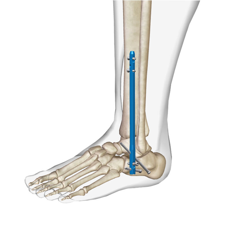
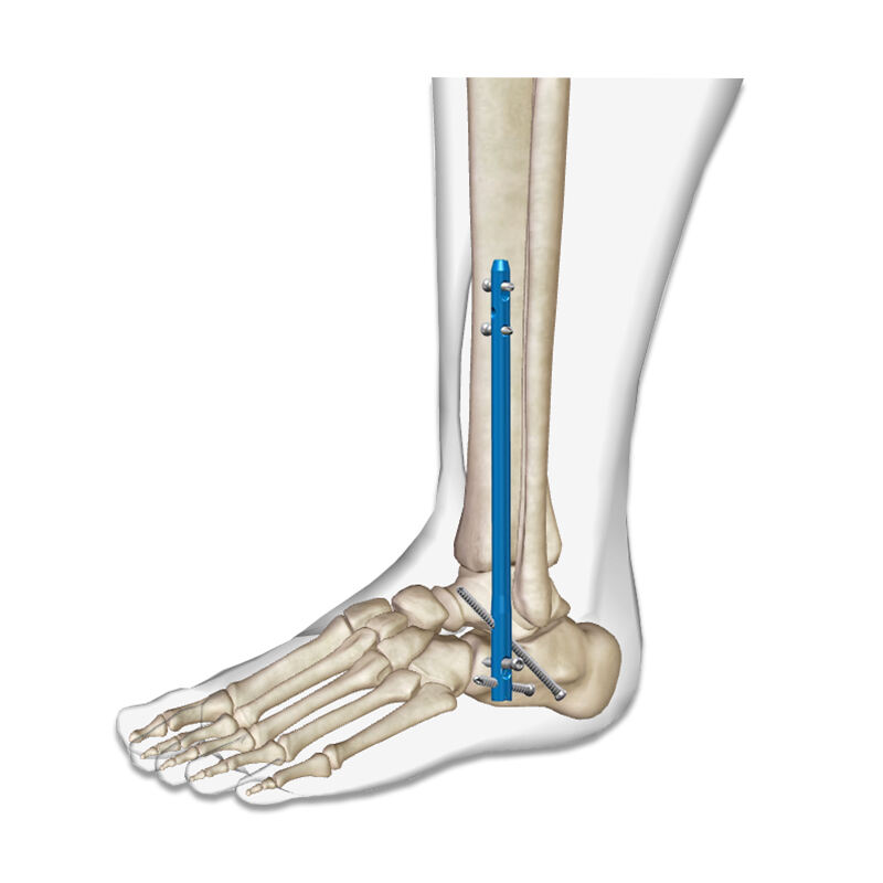
The evolution of orthopedic surgical techniques has brought significant advancements in treating complex ankle pathologies, particularly through the implementation of modern fixation systems. An intramedullary nail system represents a revolutionary approach to ankle joint fusion procedures, offering enhanced stability and improved patient outcomes compared to traditional methods. This sophisticated implant technology has transformed the landscape of ankle arthrodesis by providing superior biomechanical properties and facilitating optimal bone healing. Surgeons worldwide are increasingly adopting this innovative approach due to its proven efficacy in addressing challenging cases where conventional plate and screw fixation may fall short.
Biomechanical Advantages of Intramedullary Nail Systems
Load Distribution and Stress Management
The biomechanical superiority of an intramedullary nail system lies in its ability to distribute loads along the entire length of the implant rather than concentrating stress at specific points. This design principle creates a more physiological load transfer pattern that closely mimics natural bone mechanics. The central positioning within the medullary canal allows for optimal alignment with the mechanical axis of the limb, reducing the risk of implant failure and promoting uniform bone remodeling. Clinical studies have demonstrated that this load-sharing mechanism significantly reduces the incidence of stress shielding, a common complication associated with rigid plate fixation systems.
Furthermore, the intramedullary positioning provides enhanced resistance to bending and torsional forces that commonly occur during weight-bearing activities. The implant's geometry creates a composite structure with the surrounding bone, where both materials contribute to overall mechanical strength. This synergistic relationship is particularly beneficial in ankle fusion procedures, where the joint must withstand substantial forces during locomotion. The superior mechanical properties translate to improved implant longevity and reduced revision rates in clinical practice.
Compression and Stability Mechanisms
Modern intramedullary nail systems incorporate sophisticated compression mechanisms that enable surgeons to achieve optimal interfragmentary compression at the fusion site. The controlled compression promotes primary bone healing by maintaining intimate contact between bone surfaces while minimizing micro-motion that could impede fusion. The compression is typically achieved through specialized compression screws or expansion mechanisms within the nail itself, allowing for precise control over the compression force applied.
The multi-directional locking capability of contemporary systems provides additional rotational stability, which is crucial for successful ankle arthrodesis. The combination of axial compression and rotational control creates an ideal mechanical environment for bone fusion. This comprehensive stability allows for earlier weight-bearing in many cases, potentially accelerating the rehabilitation process and improving patient satisfaction. The enhanced stability also reduces the need for external immobilization, contributing to better functional outcomes and reduced complications associated with prolonged immobilization.
Clinical Applications and Patient Selection
Primary Ankle Arthrodesis Indications
The application of an intramedullary nail system in primary ankle arthrodesis has shown remarkable success in treating various pathological conditions. Primary indications include end-stage ankle arthritis, severe post-traumatic arthritis, avascular necrosis of the talus, and complex deformities requiring correction. The system is particularly valuable in cases where significant bone loss or poor bone quality makes traditional fixation methods inadequate. Patients with rheumatoid arthritis or other inflammatory conditions often benefit from this approach due to the superior holding power and reduced reliance on peripheral bone quality.
The versatility of the system allows for simultaneous correction of angular deformities while achieving solid fusion. This dual capability is especially important in patients with severe hindfoot malalignment, where restoration of proper mechanical axis is essential for optimal long-term outcomes. The ability to address both fusion and deformity correction in a single procedure reduces surgical complexity and potentially improves patient compliance with post-operative protocols.
Revision and Salvage Procedures
In revision scenarios, where previous fusion attempts have failed or complications have arisen, the intramedullary nail system offers significant advantages over repeat plating procedures. The central canal positioning bypasses many of the soft tissue complications associated with previous surgical approaches, reducing the risk of wound healing problems and infection. The robust fixation provided by the system is particularly valuable in cases with poor bone quality or significant bone defects resulting from previous failed surgeries.
Salvage procedures following failed total ankle replacements represent another important application area. The system can effectively bridge significant bone defects while providing stable fixation in compromised bone stock. The ability to span multiple joints when necessary makes it an excellent option for complex reconstructive procedures where traditional methods may be insufficient. These challenging cases often require creative surgical solutions, and the versatility of intramedullary nail systems provides surgeons with the flexibility needed to address complex anatomical situations.
Surgical Technique and Technical Considerations
Pre-operative Planning and Imaging
Successful implementation of an intramedullary nail system requires meticulous pre-operative planning utilizing advanced imaging modalities. Three-dimensional CT scanning provides essential information about bone quality, canal diameter, and the presence of any anatomical variants that might influence surgical approach. Weight-bearing radiographs help assess the degree of deformity and aid in determining the appropriate correction angles required during surgery. Pre-operative templating allows surgeons to select optimal nail dimensions and predict potential technical challenges before entering the operating room.
Advanced imaging also helps identify potential contraindications such as severe canal sclerosis, previous hardware that might interfere with nail placement, or anatomical variants that could complicate the procedure. The assessment of soft tissue conditions through MRI can provide valuable information about the presence of infection, osteomyelitis, or other conditions that might influence surgical timing or approach selection. Comprehensive pre-operative evaluation ensures optimal patient selection and maximizes the likelihood of successful outcomes.
Operative Technique and Nail Insertion
The surgical technique for intramedullary nail system insertion requires precise execution of multiple critical steps to ensure optimal outcomes. The procedure typically begins with careful patient positioning and the establishment of appropriate surgical approaches that provide adequate visualization while minimizing soft tissue trauma. The entry point selection is crucial, as improper positioning can lead to nail malposition and suboptimal mechanical properties. Gradual reaming of the medullary canal must be performed with careful attention to preserve cortical bone integrity while creating appropriate space for nail insertion.
Joint preparation represents a critical phase of the procedure, requiring complete removal of articular cartilage and subchondral bone to create bleeding bone surfaces conducive to fusion. The preparation must achieve flat, congruent surfaces while maintaining appropriate bone stock for stable fixation. Proper nail insertion technique involves careful attention to rotation and depth to ensure optimal positioning within the canal. The final locking sequence must be executed with precision to achieve appropriate compression while avoiding over-tightening that could compromise bone integrity or implant performance.
Post-operative Management and Rehabilitation
Immediate Post-operative Care
The post-operative management protocol following intramedullary nail system implantation requires careful balance between protecting the surgical site and promoting early mobilization. Initial management focuses on pain control, wound care, and monitoring for early complications such as infection or neurovascular compromise. The enhanced stability provided by the system often allows for earlier weight-bearing compared to traditional fixation methods, but the specific timeline must be individualized based on bone quality, patient factors, and surgical findings.
Radiographic monitoring during the early post-operative period helps assess nail position, compression maintenance, and early signs of bone healing. Serial imaging studies provide valuable feedback about the progression of fusion and can help identify potential complications before they become clinically significant. The frequency and timing of follow-up visits must be tailored to individual patient needs while ensuring adequate surveillance for complications or delayed healing.
Long-term Rehabilitation and Outcomes
The rehabilitation process following intramedullary nail system implantation typically involves a graduated progression of activity levels designed to promote bone healing while preventing complications. Physical therapy interventions focus on maintaining range of motion in adjacent joints, preventing muscle atrophy, and gradually restoring functional activities. The superior stability provided by the system often allows for more aggressive rehabilitation protocols compared to traditional fixation methods, potentially leading to faster return to activities of daily living.
Long-term outcomes with intramedullary nail systems have shown encouraging results in terms of fusion rates, patient satisfaction, and functional improvement. Studies demonstrate fusion rates consistently above 90% in most patient populations, with significant improvements in pain scores and functional outcome measures. The durability of the fixation has proven excellent in long-term follow-up studies, with low rates of implant failure or need for revision surgery. Patient-reported outcomes consistently show high satisfaction levels with the procedure and its results.
Complications and Risk Management
Intraoperative Complications
While intramedullary nail system implantation is generally considered a safe procedure, various intraoperative complications can occur and require immediate recognition and management. Cortical perforation during reaming or nail insertion represents one of the most common technical complications, potentially compromising the mechanical integrity of the construct. Careful technique and appropriate imaging guidance can minimize this risk, but surgeons must be prepared to address perforations when they occur through modified surgical techniques or alternative fixation strategies.
Neurovascular injury, though uncommon, represents a serious potential complication that requires immediate recognition and appropriate management. The proximity of important neurovascular structures to the surgical field necessitates careful anatomical awareness and gentle tissue handling throughout the procedure. Intraoperative monitoring and immediate assessment of neurovascular function can help identify problems early and facilitate prompt intervention when necessary.
Post-operative Complications and Management
Post-operative complications following intramedullary nail system implantation can range from minor issues to serious problems requiring additional intervention. Wound healing complications, while less common than with plate fixation, can still occur and may require aggressive management including debridement, antibiotic therapy, or even implant removal in severe cases. Early recognition of infection signs and prompt treatment are essential for preserving the surgical result and preventing more serious complications.
Non-union or delayed union represents another potential complication that may require additional surgical intervention. Risk factors for healing complications include smoking, diabetes, poor bone quality, and inadequate initial fixation. Management strategies may include bone grafting, revision fixation, or biological enhancement techniques depending on the specific circumstances. Regular monitoring and early intervention when problems are identified can often salvage difficult cases and achieve successful outcomes.
Future Developments and Innovations
Material Science Advancements
The future of intramedullary nail system technology lies in continued advancement of materials science and implant design. New titanium alloys and surface treatments are being developed to enhance osseointegration and reduce the risk of implant-related complications. Bioactive coatings and drug-eluting surfaces show promise for reducing infection rates and promoting faster bone healing. These innovations may lead to improved fusion rates and reduced complications in challenging patient populations.
Advanced manufacturing techniques including 3D printing and patient-specific implant design are opening new possibilities for customized treatment approaches. These technologies may allow for better anatomical fit and improved mechanical properties tailored to individual patient anatomy. The integration of smart materials and sensors into implant design could provide real-time feedback about healing progress and mechanical loading, potentially revolutionizing post-operative monitoring and rehabilitation protocols.
Surgical Technology Integration
The integration of advanced surgical technologies with intramedullary nail system procedures continues to evolve, offering improved precision and outcomes. Computer-assisted navigation systems provide enhanced accuracy in nail placement and alignment correction, potentially reducing complications and improving long-term results. Robotic assistance in surgery may further enhance precision while reducing surgeon fatigue and variability in technique execution.
Advanced imaging technologies including intraoperative CT and fluoroscopy integration are improving real-time visualization and decision-making during surgery. These technologies allow for immediate assessment of nail position, compression achievement, and alignment correction, enabling surgeons to make intraoperative adjustments that optimize outcomes. The continued development of minimally invasive techniques may further reduce surgical trauma while maintaining the excellent mechanical properties of intramedullary nail systems.
FAQ
What makes intramedullary nail systems superior to traditional plate fixation for ankle fusion?
Intramedullary nail systems offer several key advantages including better load distribution along the entire implant length, enhanced rotational stability, and reduced soft tissue complications. The central canal positioning provides superior biomechanical properties and allows for earlier weight-bearing compared to peripheral plate fixation. Additionally, the compression mechanisms enable optimal interfragmentary compression for improved fusion rates.
How long does the recovery process typically take after intramedullary nail system implantation?
Recovery timelines vary based on individual factors, but most patients can expect initial healing within 6-8 weeks, with progressive weight-bearing allowed earlier than traditional methods due to enhanced stability. Complete bone fusion typically occurs within 3-6 months, though this can vary depending on patient factors such as age, bone quality, and compliance with post-operative restrictions. Full functional recovery may take 6-12 months depending on individual circumstances.
Are there specific contraindications for intramedullary nail system use in ankle fusion?
Contraindications may include active infection at the surgical site, severe canal sclerosis preventing nail insertion, inadequate bone stock for stable fixation, or certain anatomical variants that make nail placement technically impossible. Relative contraindications include severe osteoporosis, significant medical comorbidities affecting healing, or patient factors that may compromise compliance with post-operative protocols.
What are the long-term outcomes and implant durability expectations?
Long-term studies demonstrate excellent outcomes with fusion rates typically exceeding 90% and high patient satisfaction scores. The durability of modern intramedullary nail systems is excellent, with low rates of implant failure or need for revision surgery over 10-15 year follow-up periods. Most patients experience significant pain relief and functional improvement that is maintained long-term, though some activity modifications may be necessary to optimize implant longevity.
Table of Contents
- Biomechanical Advantages of Intramedullary Nail Systems
- Clinical Applications and Patient Selection
- Surgical Technique and Technical Considerations
- Post-operative Management and Rehabilitation
- Complications and Risk Management
- Future Developments and Innovations
-
FAQ
- What makes intramedullary nail systems superior to traditional plate fixation for ankle fusion?
- How long does the recovery process typically take after intramedullary nail system implantation?
- Are there specific contraindications for intramedullary nail system use in ankle fusion?
- What are the long-term outcomes and implant durability expectations?
