EN


How to Accurately Position the PHILOS Plate in Proximal Humeral Fractures: The “K-Wire Positioning Technique”
For Neer type III and IV proximal humeral fractures, head-preserving strategies mainly include the PHILOS plate and intramedullary nailing. The PHILOS plate is specifically designed for proximal humeral fractures. Locking screws inserted at different angles provide three-dimensional purchase of the proximal humeral fragments, and calcar screws can effectively prevent varus collapse. Clinical experience over many years has confirmed reliable outcomes.
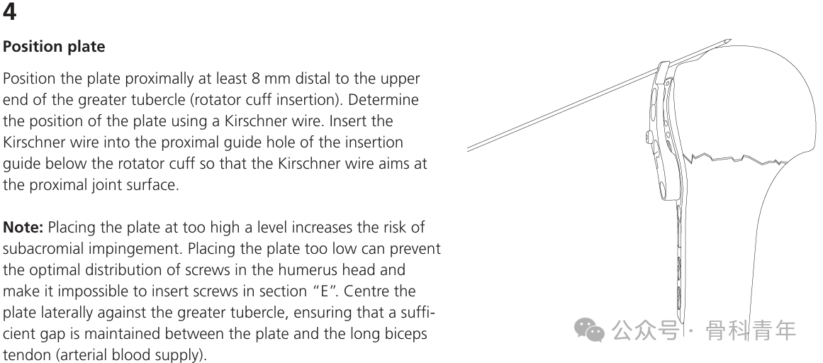
Theoretically, the ideal position of the PHILOS plate is 2–4 mm posterior to the bicipital groove and 5–8 mm below the apex of the greater tuberosity.
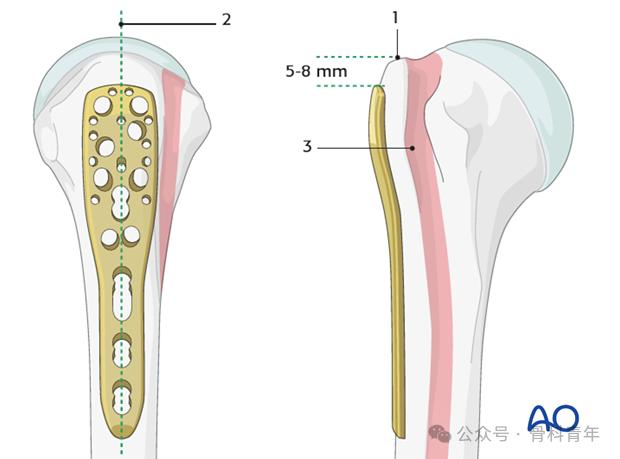
▲ Standard position of the PHILOS plate in the proximal humerus. Source: AO Surgery Reference.
However, in clinical practice, after fracture reduction and temporary fixation, accurate judgment of PHILOS plate position often takes considerable time and frequently requires repeated temporary K-wire fixation and fluoroscopic adjustment. This is especially true for those of us in China who habitually work without lead aprons and stand outside the operating room behind a lead screen during fluoroscopy. Repeated fluoroscopic adjustment undoubtedly prolongs operative time and increases the risk of surgical infection.
To accurately position the PHILOS plate, orthopaedic surgeons from the First Hospital of Longyan City, Fujian Province, adopted a “K-wire positioning technique,” which effectively addresses this problem. This issue shares the technique for reference by orthopaedic surgeons (reference at end).

Technique of the “K-Wire Positioning Technique”
After routine disinfection and draping, a deltopectoral approach is used. Following reduction and temporary fixation of the proximal humerus, a single 2.0-mm K-wire is used as a reference for plate placement.
Specific steps: 3-mm mini-incision is made above the greater tuberosity along the fibers of the supraspinatus tendon (a miniaturized version of the rotator cuff incision used in humeral intramedullary nailing). The blunt end of the 2.0-mm K-wire is gently introduced through the supraspinatus mini-incision until it contacts the articular cartilage surface of the humeral head. Care is taken not to penetrate the cartilage, injure the humeral head cartilage, or enter the glenohumeral joint. The skin insertion depth of the K-wire is generally 2–3 cm, and the blunt tip is kept outside the joint cavity throughout. The PHILOS plate is placed against the greater tuberosity, immediately beneath the lower edge of the K-wire:
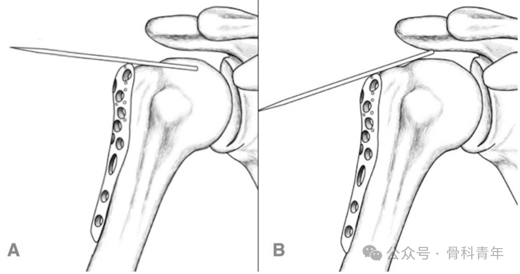
▲ Figure A: The blunt end of a 2.0-mm K-wire is gently introduced through a mini-incision in the supraspinatus and contacts the articular cartilage surface of the humeral head (without penetrating the cartilage); the PHILOS plate is placed against the greater tuberosity and aligned with the lower edge of the K-wire. B: The K-wire is rotated back and forth; the blunt tip slides along the articular surface of the humeral head. When the blunt tip slides past the highest point of the humeral head cartilage, it tilts upward, causing the sharp end of the K-wire to move downward. The downward-moving sharp end presses the PHILOS plate downward, gradually lowering the plate height.
Using the 3-mm supraspinatus tendon incision as a fulcrum for rotation, the K-wire is rotated back and forth; the blunt tip slides along the articular surface of the humeral head. When the blunt tip slides past the highest point of the humeral head cartilage, the blunt tip tilts upward, and the sharp end of the K-wire moves downward accordingly. The downward-moving sharp end presses the PHILOS plate downward, lowering the plate placement height. After multiple to-and-fro rotations, the K-wire is used to gradually push the plate downward. The lowest position that the plate can reach during rotation is recorded intraoperatively and used as the reference landmark for the apex of the greater tuberosity. Under direct vision, the plate is placed 2–4 mm below the lower edge of the K-wire (approximately 1–2 diameters of a 2.0-mm K-wire). Fluoroscopy confirms satisfactory position of the proximal humeral plate, after which an adequate number of locking screws are inserted proximally and distally.
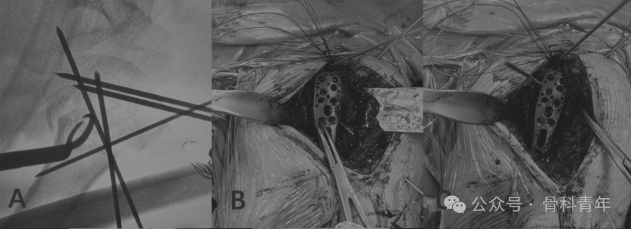
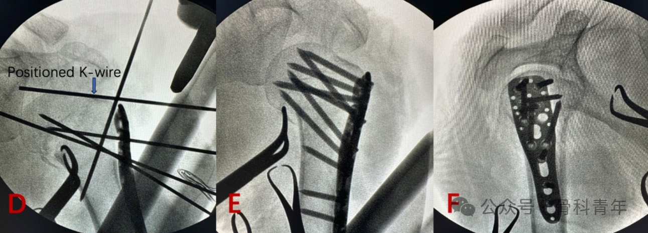
▲ Intraoperative steps of K-wire-assisted positioning during PHILOS plate placement in a 65-year-old woman with a Neer three-part proximal humeral fracture. A: Anatomic reduction completed with crossed temporary K-wire fixation. B: The blunt end of a 2.0-mm K-wire is directed obliquely against the area above the apex of the greater tuberosity and points toward the highest point of the spherical humeral head; the exposed extracorporeal segment of the K-wire serves as a linear reference for the apex of the greater tuberosity. The PHILOS plate is aligned with the lower edge of the K-wire and placed 2–4 mm below it (approximately 1–2 diameters of a 2.0-mm K-wire under direct vision). C, D: The plate is temporarily fixed with K-wires, and fluoroscopy confirms satisfactory plate position. E, F: Final screws are inserted, and fluoroscopy from multiple angles confirms screw distribution, length, and position.
Study results showed that this positioning technique effectively reduced intraoperative fluoroscopy frequency, shortened operative time, and decreased intraoperative blood loss.
In essence, this technique is consistent with the instrument-based plate positioning guide, except that we do not routinely use it or do not have it available.
Que, W., Tu, X., Xie, J. et al. Kirschner wire-assisted positioning technique reduces operative time and fluoroscopy exposure in proximal humeral PHILOS plate placement: a retrospective comparative study. J Orthop Surg Res21, 349 (2026).