EN


Two Internal Fixation Methods for Clavicle Fractures: Techniques, Tips, and Pitfalls
Clavicle fractures are among the most common fractures. Surgical internal fixation is usually performed with plates or intramedullary nails. Today we summarize the relevant technical points.
To operate or not? When to operate?
Surgery is not omnipotent, but choosing the wrong indication is fatal.
- Initial decision-making (the Murray model)
If every displaced fracture were operated on, the number needed to treat (NNT) to prevent one nonunion would be 7.5. However, if the patient is a smoker with a comminuted fracture and severe displacement, the nonunion probability reaches 40% — here surgery must be performed decisively, and the NNT drops sharply to 1.7.
- The 6-week "reconsideration window" (the Nicholson gold standard)
For patients initially treated conservatively, assessment must be carried out at week 6. Look for the following three findings:
① QuickDASH score of 40;
② no bridging callus on X-ray or ultrasound;
③ abnormal mobility on physical examination.
If any two of these are present, the nonunion rate soars to 60%, and conservative treatment must be terminated immediately in favor of surgical internal fixation.
I. Plate Fixation: The Trade-off Between Stability and Soft Tissue
The challenge with plating lies not in inserting screws, but in preserving local blood supply and avoiding danger zones.
Tips and Tricks
1. Completely abandon the 3.5 mm reconstruction plate.
Never compromise biomechanics for the sake of "easy contouring"! The literature confirms that reconstruction plates have a fatigue-failure and mechanical-failure rate of up to 53% in complex comminuted fractures. Instead, use an anatomically contoured locking compression plate (ALCP), which offers twice the yield strength, or a biomechanically superior dual mini-plate construct (superior 2.7 mm + anterior 2.4 mm).
2. A subtle change in positioning to buy a lifeline.
The beach-chair position is strongly recommended. Studies confirm that, compared with the supine position, the beach-chair position alters soft-tissue tension and increases the safe distance between the neurovascular bundle and the drill at the danger zone by an additional 1.4 mm.
3. A decisive edge in plate positioning.
Superior plating is strong against compression but is highly prone to soft-tissue irritation (implant removal rate 20%–50%).
Antero-inferior plating is strongly recommended: it sits hidden beneath the pectoralis major and deltoid fascia, completely resolving irritation pain; furthermore, the antero-inferior cantilever has excellent bending stiffness, with less intraoperative blood loss.
4. MIPO and the bridging concept. For butterfly fragments, insisting on anatomical reduction is simply manufacturing a nonunion. Minimally invasive plate osteosynthesis (MIPO) should be used to create an extraperiosteal tunnel. This not only reduces the incidence of postoperative anterior chest numbness from 55% to 12.5%, but also maximally preserves the fracture-site hematoma and promotes healing.
Practical Pitfalls and How to Avoid Them
1. The drill's danger zones
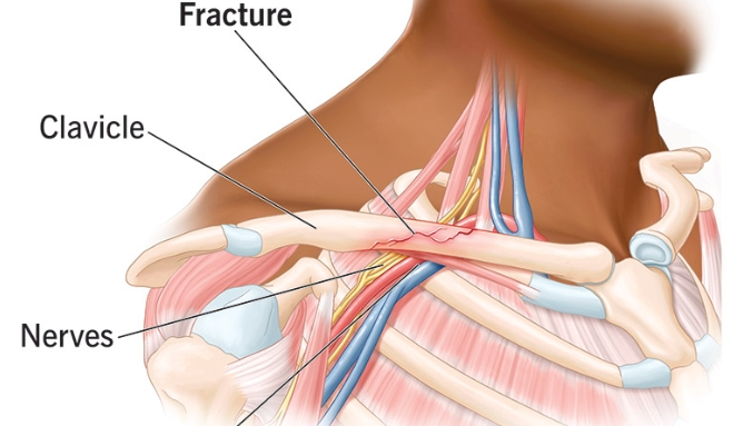
Medial third (extreme-risk zone). The subclavian vein lies flush against the posterior cortex (on average only 4.77 mm, and in some cases 0 mm). With an antero-inferior plate, screws here must be directed from anterior to posterior. Pitfall avoidance: the most medial screw should be unicortical only, or the drill hole should be deliberately angled superolaterally — never drive straight through!
Middle third (high-risk zone). The neurovascular bundle lies posteroinferiorly. With superior plating, drilling downward points directly at the artery and vein. Pitfall avoidance: a mechanical depth limiter (set at 15–20 mm) must be used, and a blunt retractor should be placed beneath the cortex to protect the subclavius muscle.
Lateral third (safe zone). The vascular plexus descends into the axilla, well beyond 60 mm from the cortex; here one can operate confidently and without hesitation.
2. The "refracture" curse after hardware removal
Large plates cause stress shielding; once removed, the empty screw holes are fatal stress risers. Data show that the refracture rate within 2 months of plate removal is as high as approximately 9%. Preoperatively, it must be clearly communicated that the plate should not be removed unless necessary; strictly immobilize postoperatively.
II. Intramedullary Fixation (TENs/ESIN): The Art of Minimally Invasive Surgery and Biomechanical Compromise
Elastic nails provide relative stability and heal through callus formation (on average 1–3 weeks faster than plates), with a small incision and a 0% refracture rate after removal — but when applied to the wrong indication, they are a disaster.
Tips and Tricks
1. Strictly observe the indication red line. Suitable only for Robinson type 2B1 fractures (simple transverse or short oblique). Absolutely contraindicated for comminuted (OTA 15-B2/B3) or length-unstable fractures.
2. The golden entry point. Enter the anterior cortex 1.5–2.0 cm lateral to the sternoclavicular joint (sternal entry), avoiding the acromioclavicular joint injury caused by lateral entry.
3. Cut down decisively. If closed reduction (joystick technique) does not go smoothly, immediately convert to a mini-open approach (1.5 cm) for direct visualization and reduction. Never resort to forceful blind passage, which risks puncturing the pleural cupola above the first rib and causing iatrogenic pneumothorax.
Practical Pitfalls and How to Avoid Them
1. Extremely poor rotational control and telescoping. A single smooth titanium nail cannot control rotation or axial compression. Pitfall avoidance: during the first 3–4 postoperative weeks, strict immobilization in a sling is mandatory; abduction or forward flexion of the shoulder beyond 90° is absolutely prohibited until soft callus has formed.
2. Nail migration and medial skin breakdown (the most common complication). Migration of the titanium nail is an early nightmare. Pitfall avoidance: modern surgery must mandatorily use "terminal end caps"! Locking the end against the medial cortex completely solves the problems of nail migration and skin irritation.
Summary: For complex comminuted fractures, choose dual plates or anatomical plates — remember antero-inferior placement and bridging MIPO to avoid vascular injury. For simple transverse fractures, choose elastic intramedullary nails — remember that end caps are mandatory and strict immobilization is required to prevent nail migration.