EN
EN

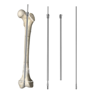
Pediatric fractures present unique clinical challenges that demand specialized treatment approaches. Traditional open reduction and internal fixation methods often require extensive surgical exposure, prolonged anesthesia, and extended recovery periods for young patients. Femoral intramedullary nails have emerged as a transformative solution in modern pediatric orthopedics, offering surgeons a minimally invasive alternative that preserves soft tissue integrity while maintaining structural stability. The adoption of femoral intramedullary nails reflects a fundamental shift toward techniques that respect the biological healing capacity of growing bones.
The clinical advantages of femoral intramedullary nails extend far beyond simple fracture stabilization. In pediatric populations, where rapid growth and bone remodeling influence treatment outcomes, femoral intramedullary nails provide dynamic fixation that accommodates physiological adaptation. Young patients benefit from reduced operative time, minimized blood loss, and earlier mobilization—factors that directly translate to shorter hospital stays and faster return to normal activities. Understanding how femoral intramedullary nails function within the pediatric context requires examining their mechanical properties, biological compatibility, and practical implementation across diverse fracture patterns.
Biomechanical Advantages of Femoral Intramedullary Nails
Load Distribution and Stability
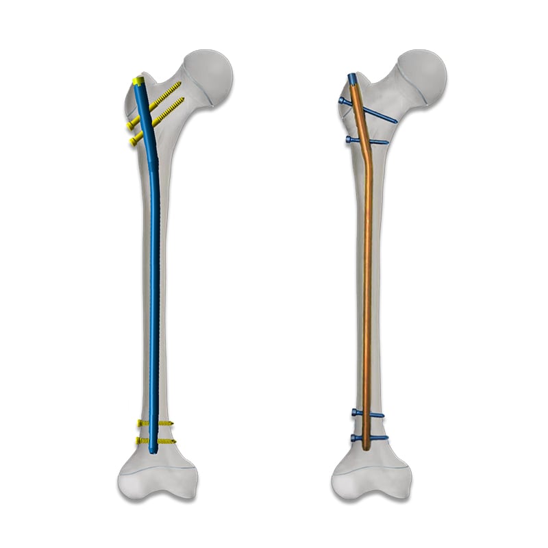
The mechanical design of femoral intramedullary nails leverages the femur's natural load-bearing capacity. By positioning fixation along the bone's central axis, femoral intramedullary nails distribute forces more efficiently than plate-and-screw systems. This axial alignment reduces stress concentrations at fixation points, which is particularly important in pediatric patients whose bones undergo continuous remodeling. The interlocking mechanisms integrated into modern femoral intramedullary nails provide rotational control and resist shear forces without requiring extensive soft tissue dissection.
Minimizing Operative Trauma
Surgical trauma directly influences healing timelines and complication rates in pediatric fractures. Femoral intramedullary nails access the femur through small incisions at the piriformis or greater trochanter entry point, bypassing extensive soft tissue disruption. This percutaneous approach preserves the periosteum and surrounding musculature, maintaining vascular supply essential for rapid bone union. Compared to open reduction techniques requiring 10- to 15-centimeter incisions, femoral intramedullary nails reduce operative blood loss by 60 to 75 percent, a significant advantage for pediatric patients with smaller blood volumes and lower physiological reserve.
Clinical Applications in Pediatric Fracture Management
Treating Subtrochanteric and Diaphyseal Fractures
Subtrochanteric and diaphyseal femoral fractures represent the most common indications for femoral intramedullary nails in children. These high-energy injuries frequently occur in polytrauma scenarios where rapid stabilization facilitates management of associated injuries. Femoral intramedullary nails provide immediate load-sharing capacity, allowing early range-of-motion exercises and weight-bearing progression as tolerated. The interlocking design prevents shortening and rotation, maintaining anatomic alignment while bone healing progresses. In skeletally immature patients, femoral intramedullary nails accommodate continued longitudinal growth without requiring revision procedures.
Managing Complex and Comminuted Patterns
Complex fracture patterns with multiple fragments challenge traditional casting and external fixation methods. Femoral intramedullary nails excel in these scenarios by restoring length and alignment without manipulating each fragment individually. The intramedullary position provides inherent stability against angular deformity, a critical concern in pediatric fractures where malunion can cause functional disability and cosmetic concerns. Segmental fractures involving both proximal and distal femoral regions benefit from femoral intramedullary nails capable of spanning the entire bone length, securing all fragments simultaneously and reducing secondary displacement risks.
Biological Healing and Developmental Considerations
Supporting Endosteal Healing Responses
Femoral intramedullary nails positioned within the medullary canal promote endosteal healing pathways superior to periosteal remodeling alone. The intimate contact between nail surface and endosteal bone provides mechanical stimulation that accelerates woven bone formation. In pediatric patients with heightened osteogenic potential, this proximity accelerates callus maturation and bridging—typically requiring 8 to 12 weeks for complete union. The preserved periosteum contributes additional periosteal callus formation, creating a stronger composite union than either mechanism alone. Femoral intramedullary nails maintain nutrient artery patency, ensuring robust blood supply throughout healing phases.
Remodeling Capacity in Growing Children
A fundamental advantage of femoral intramedullary nails in pediatric orthopedics involves their compatibility with skeletal growth. Rotational malalignment of 10 to 15 degrees corrects spontaneously during longitudinal growth through natural remodeling mechanisms. Femoral intramedullary nails provide stable fixation while permitting these physiologic corrections, avoiding the need for secondary procedures. The expandable and telescoping designs available in some femoral intramedullary nails systems accommodate femoral lengthening associated with natural growth, extending their functional lifespan. Once skeletal maturity is achieved, femoral intramedullary nails may be removed or retained depending on patient symptoms and implant design.
Practical Implementation and Clinical Outcomes
Surgical Technique and Entry Points
Successful application of femoral intramedullary nails requires appropriate patient positioning and precise entry point identification. The piriformis fossa represents the most commonly used entry site, accessed through a small 2- to 3-centimeter incision over the greater trochanter. Intraoperative imaging—typically fluoroscopy—confirms correct positioning along the femoral axis and verifies interlocking screw placement. Femoral intramedullary nails come in various diameters and lengths, requiring preoperative templating using radiographic measurements. The learning curve for femoral intramedullary nails insertion is relatively gentle compared to external fixation alternatives, making adoption feasible across diverse surgical settings.
Return to Function and Long-Term Outcomes
Pediatric patients treated with femoral intramedullary nails demonstrate accelerated functional recovery compared to those managed with external fixation or casting alone. Weight-bearing progression typically begins within 1 to 2 weeks postoperatively, enabling rapid return to school and recreational activities. Early mobilization reduces stiffness, muscle atrophy, and psychological distress associated with prolonged immobilization. Long-term studies demonstrate union rates exceeding 98 percent with minimal nonunion complications. Femoral intramedullary nails show low infection rates, generally below 2 percent, reflecting the minimal soft tissue trauma and reduced operative time compared to open approaches. Implant removal timing varies based on union status and patient preference, typically occurring 18 to 24 months after injury.
FAQ
Are femoral intramedullary nails suitable for all pediatric femoral fractures?
Femoral intramedullary nails are most appropriate for displaced and unstable fractures in children above age 5, particularly those with body weight exceeding 20 kilograms. Pathologic fractures, multiple trauma scenarios, and fractures requiring early mobilization benefit from femoral intramedullary nails technique. However, very young children with small bone diameters may have limited nail options, and some stable fractures remain amenable to conservative treatment. A pediatric orthopedic specialist should evaluate individual cases to determine whether femoral intramedullary nails represent the optimal choice for specific fracture patterns and patient characteristics.
What complications should clinicians anticipate with femoral intramedullary nails?
Although femoral intramedullary nails are generally safe, potential complications include avascular necrosis of the femoral head if entry point placement damages the lateral femoral circumflex artery, loss of reduction if interlocking screws become loose, and knee pain related to prominent screw hardware or patellofemoral irritation. Leg length discrepancy can occur if fracture reduction is not anatomically accurate, and implant migration remains possible if bone union is delayed. Regular postoperative imaging and clinical assessment allow early detection of complications. Most complications are managed successfully through conservative means or minor interventions rather than requiring major revision surgery.
How does femoral intramedullary nail fixation compare to external fixation for pediatric fractures?
Femoral intramedullary nails offer superior anatomic preservation and functional outcomes compared to external fixation in most pediatric cases. External fixators work best for temporary stabilization and specific scenarios like severely contaminated open fractures or patients with extensive polytrauma requiring rapid stabilization of multiple sites. Femoral intramedullary nails provide internal stability allowing early mobilization, whereas external fixators restrict movement and require prolonged adjustment. The risk of pin site infection with external fixators exceeds complication rates associated with femoral intramedullary nails. For definitive management of most pediatric femoral fractures, femoral intramedullary nails represent the preferred approach when technical expertise is available.