EN
EN

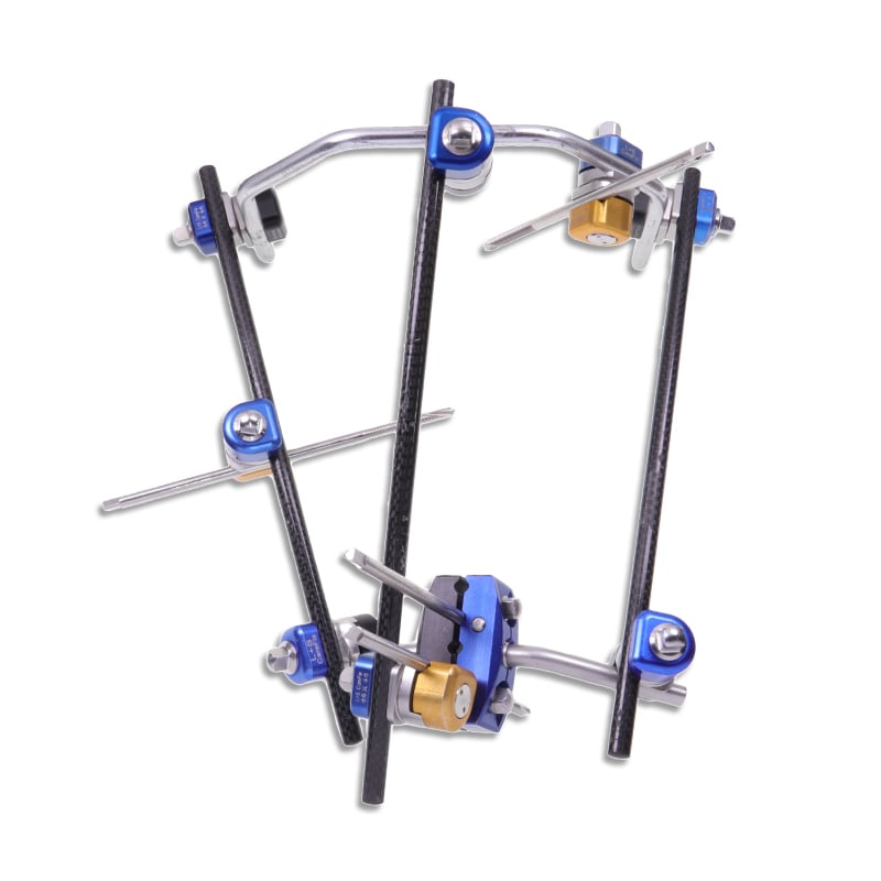
Modern orthopedic surgery increasingly demands solutions that minimize tissue trauma while maintaining robust fracture stabilization. External fixation has emerged as a cornerstone technology for achieving this balance, particularly in complex fracture management where traditional open reduction may compromise soft tissue integrity or patient outcomes. The evolution of external fixation systems now enables surgeons to deploy multi-mode capabilities that adapt to diverse clinical scenarios, from simple fracture reduction to complex limb reconstruction procedures. Understanding how external fixation facilitates minimally invasive treatment requires examining both the mechanical principles and the clinical advantages that distinguish it from alternative fixation methods.
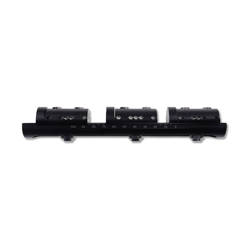
External fixation represents a departure from conventional plate-and-screw fixation by positioning the stabilizing apparatus entirely outside the fracture site. This positioning allows surgeons to reduce and stabilize complex fractures without extensive soft tissue disruption, preserving blood supply and reducing infection risk. The multi-mode nature of contemporary external fixation systems means surgeons can select from unilateral, bilateral, or hybrid configurations depending on fracture geometry, patient anatomy, and healing requirements. This flexibility transforms external fixation from a trauma-only tool into a precision instrument suitable for elective orthopedic procedures where minimally invasive treatment principles guide clinical decision-making.
Mechanical Advantages of External Fixation in Fracture Stabilization
Load Distribution and Structural Rigidity
The biomechanical foundation of external fixation relies on positioning fixation elements—pins, half-pins, or tensioned wires—around the fracture site rather than directly across it. This geometry allows external fixation to distribute loads through multiple vectors, creating highly rigid constructs that rival or exceed the stiffness of internal implants. When properly configured, external fixation provides three-dimensional stability that prevents shear displacement, rotation, and axial collapse, all critical factors in early mobilization and fracture healing. The modular nature of modern external fixation systems permits surgeons to adjust the configuration intraoperatively if initial assessment reveals unexpected instability patterns or comminution severity.
Graduated Load Transfer for Biological Healing
Unlike rigid internal fixation, external fixation systems can be progressively dynamized to transfer increasing loads to the healing bone. This graduated load transfer stimulates osteogenic response and promotes secondary bone formation, potentially accelerating healing in certain fracture patterns. Surgeons can adjust external fixation frames during follow-up visits without additional surgery, fine-tuning mechanical conditions as the fracture callus develops and matures. This biological advantage explains why external fixation frequently produces superior functional outcomes in periosteal-rich fractures and pediatric cases where biological healing potential remains high.
Clinical Applications in Minimally Invasive Treatment Paradigms
Complex Fracture Reduction Without Soft Tissue Compromise
Minimally invasive fracture treatment prioritizes soft tissue preservation, infection prevention, and rapid return to function. External fixation excels in this context because reduction and stabilization occur through percutaneous pin insertion rather than extensile surgical approaches. In pilon fractures, plateau fractures, and complex metaphyseal injuries, external fixation preserves the periosteal blood supply while allowing excellent visualization for anatomic reduction. The multi-mode capabilities of advanced external fixation systems enable surgeons to employ techniques like ligamentotaxis—gradual reduction through traction—to achieve acceptable alignment without direct visualization of the fracture site, exemplifying true minimally invasive methodology.
Staged Treatment Protocols and Frame Conversion
Many modern external fixation protocols employ staged treatment where external fixation provides initial stabilization and reduction, followed by conversion to internal fixation once soft tissues recover. This two-stage approach combines the acute benefits of external fixation—rapid stabilization with minimal trauma—with the long-term advantages of internal fixation, such as improved comfort and earlier weight-bearing progression. External fixation serves as a bridge during the critical healing window when internal fixation might introduce infection risk or compromise vascular integrity. The ease of frame adjustment and repositioning during staged treatment makes external fixation the preferred choice for polytrauma patients requiring phased operative intervention.
Multi-Mode Fixation Strategies and Clinical Customization
Unilateral, Bilateral, and Hybrid Configuration Selection
The sophistication of contemporary external fixation lies in its configurational flexibility. Unilateral external fixation provides excellent visualization and ease of use for simple diaphyseal fractures where stability requirements remain moderate. Bilateral external fixation dramatically increases construct stiffness for highly comminuted fractures or when managing severe soft tissue injury alongside fracture trauma. Hybrid external fixation—combining external frames with minimal percutaneous internal implants—offers maximum stiffness while maintaining minimally invasive principles, particularly valuable in complex intra-articular injuries requiring anatomic reduction. Surgeons select configuration based on comminution pattern, fracture location, bone quality, and soft tissue status, personalizing external fixation to individual patient circumstances.
Integration with Limb Reconstruction Principles
Advanced external fixation systems enable sophisticated limb reconstruction procedures including deformity correction, bone transport, and gradual distraction osteogenesis. These applications extend external fixation beyond acute fracture treatment into elective orthopedic territory where minimally invasive principles become particularly valuable. Unilateral external fixators specifically designed for limb reconstruction allow precise control of spatial relationships, essential for correcting complex three-dimensional deformities without open surgery. The multi-mode nature of reconstruction-capable external fixation systems supports both acute fracture fixation and chronic reconstruction, eliminating the need for device changes during extended treatment courses.
Clinical Outcomes and Soft Tissue Advantages
Infection Prevention Through Tissue Preservation
Minimally invasive external fixation reduces operative time, minimizes blood loss, and critically, preserves soft tissue viability. These factors directly correlate with infection prevention, a paramount concern in orthopedic trauma. By avoiding extensile incisions that compromise periosteal perfusion and muscle vascularity, external fixation maintains natural immune responses and bacterial clearance mechanisms at the fracture site. The pin sites, while requiring diligent patient care and clinical monitoring, pose lower infection risk than the composite surgical incisions required for open reduction and internal plate fixation. In contaminated or highly comminuted fractures where infection risk escalates substantially, external fixation's tissue-sparing approach becomes particularly advantageous.
Early Mobilization and Functional Recovery
The immediate stability provided by external fixation enables early range-of-motion exercises and progressive weight-bearing, critical factors in preventing joint stiffness and improving functional recovery. Patients managed with minimally invasive external fixation typically achieve better joint mobility and earlier return to activities compared to those requiring extensive soft tissue handling. Pain reduction following less traumatic surgery further facilitates early mobilization compliance, creating a virtuous cycle where improved early function translates into superior long-term outcomes. This functional advantage explains why external fixation remains the standard of care in major trauma centers managing complex fracture patterns requiring urgent stabilization.
FAQ
How does external fixation compare to internal plate fixation for complex fractures?
External fixation preserves soft tissues and permits staged treatment, while internal plate fixation offers superior comfort and earlier weight-bearing in lower-energy injuries. The choice depends on fracture severity, soft tissue status, and contamination risk. For highly comminuted or contaminated fractures, external fixation provides superior outcomes; for simple fractures with good soft tissue cover, internal fixation may offer better patient satisfaction. Many surgeons now employ staged protocols combining both techniques, leveraging external fixation's minimally invasive advantages during acute phases.
What complications should clinicians anticipate with external fixation treatment?
Pin site infection remains the most common complication, managed through meticulous pin care education and clinical surveillance. Malunion, nonunion, and stiffness can occur if external fixation parameters are not optimized during treatment. Nerve or vessel injury during percutaneous pin insertion requires anatomic knowledge and fluoroscopic guidance. Most complications resolve with appropriate external fixation adjustment, pin care modifications, or staged conversion to internal fixation. Complication rates decrease substantially with surgeon experience and patient compliance with pin care protocols.
Can external fixation enable satisfactory outcomes in intra-articular fractures?
Yes, when combined with percutaneous reduction techniques and supplemental minimal internal fixation. Hybrid external fixation—combining external frames with small percutaneous implants—allows anatomic reduction of intra-articular fracture patterns while maintaining minimally invasive principles. Some intra-articular fractures, particularly plateau and pilon injuries, achieve excellent results through ligamentotaxis alone using external fixation, avoiding open arthrotomy. Surgeon experience with advanced reduction techniques and appropriate external fixation configuration selection determine outcomes in this challenging patient population.