EN


Proximal Femoral Intramedullary Nailing: Five Pitfalls!
Many colleagues regard intramedullary nailing for proximal femoral fractures as an entry-level procedure, but in fact “the threshold lies within the door”; the more one performs it, the more one appreciates its depth. Many postoperative complications—such as cut-out, iatrogenic fracture, and anterior cortical impingement—can actually be anticipated and avoided intraoperatively.
I. The Greatest Enemy: Reduction Pitfalls
First, it must be emphasized: reduction quality is the first determinant of success. No implant can compensate for poor reduction.
1. The Fatal “Varus Malreduction”
- Pitfall:Varus reduction (neck-shaft angle <125°) is the strongest predictor of screw cut-out.
-
Hidden killer—“Wedge Effect”:Even if reduction appears acceptable, during insertion of the main nail, because the proximal nail diameter is relatively large, inadequate reaming or a laterally placed entry point can cause the nail to act like a wedge and push the proximal fragment (femoral head-neck) into varus.
-
Dr. Wang’s pearls:
- Better slightly medial than lateral at the entry point:The entry point must be at the greater trochanter apex or slightly medial.
- Adequate reaming:Especially proximally—ream adequately; do not hesitate over that step.
- Use a hollow reamer:Compared with a solid reamer, a hollow reamer removes bone rather than compacting it, reducing displacement forces at the fracture site.
2. Easily Overlooked “Rotation” and “Distraction”
- Pitfall:On the traction table, the limb tends to externally rotate under gravity, leading to loss of postoperative anteversion (toe-out deformity); excessive traction causes fracture-site distraction (3 mm), resulting in nonunion.
-
Recommendations:
- Look at the lesser trochanter:On AP fluoroscopy, if the lesser trochanter on the affected side is much more prominent than on the contralateral side, external rotation is present and must be corrected.
- Back-tap:Before distal locking, release traction and even gently back-tap the nail or compress the fracture site (an advantage of InterTAN) to eliminate the gap.
3. Bailout Techniques for Difficult Reductions
Do not be dogmatic about closed reduction. For difficult fractures (e.g., irreducible A3 fractures):
- Joystick technique:Percutaneously insert a Schanz pin (4.0 or 5.0 mm) into the proximal femoral neck and use it like a joystick to control the proximal fragment and match it to the distal fragment, correcting flexion and varus.
- Do not hesitate:If closed reduction is poor, perform a small lateral incision (mini-open) and use reduction forceps or cerclage wire to assist reduction. This is far better than repeated fluoroscopy/radiation exposure and eventually forcing nail insertion.
II. The Entry Point Game
- Consensus for PFNA and InterTAN: The entry point is at the greater trochanter apex or slightly medial.
- Cost of lateral placement: Inevitably leads to varus reduction.
- Risk of medial placement: Injury to branches of the lateral circumflex femoral artery or iatrogenic femoral neck fracture.
- Technical detail: Adduct the affected limb 10–15° to make the greater trochanter prominent laterally, facilitating identification of the correct entry point and preventing the drill from slipping laterally.
III. Distal Impingement and Anterior Cortical Penetration
This is an anatomic trap, particularly common in Asian populations or shorter patients.
- Mechanism:The human femur has an anterior bow (mean radius of curvature [ROC] ~1200 mm), whereas older-generation main nails are straighter (ROC 1500–2000 mm). A straight nail in a curved canal will inevitably impinge on the anterior cortex distally.
- Consequences:Postoperative anterior thigh pain and even delayed femoral shaft fracture.
-
Dr. Wang’s pearls (Poller Pin technique):
- If the preoperative lateral radiograph shows a large femoral anterior bow, preplace a blocking pin (Poller pin/Steinmann pin) before reaming.
- Key position:Literature indicates that a K-wire can be placed near the anterior cortex (anterior rebar) to force the reamer and main nail posteriorly; alternatively, use the blocking principle to correct the specific direction of deviation.
- Nail selection strategy:For shorter patients or those with a large anterior bow, preferentially select a main nail with a smaller ROC (more bowed, e.g., 1.0 m or 1.5 m), or use a short nail (although short nails carry a risk of subtrochanteric stress concentration).
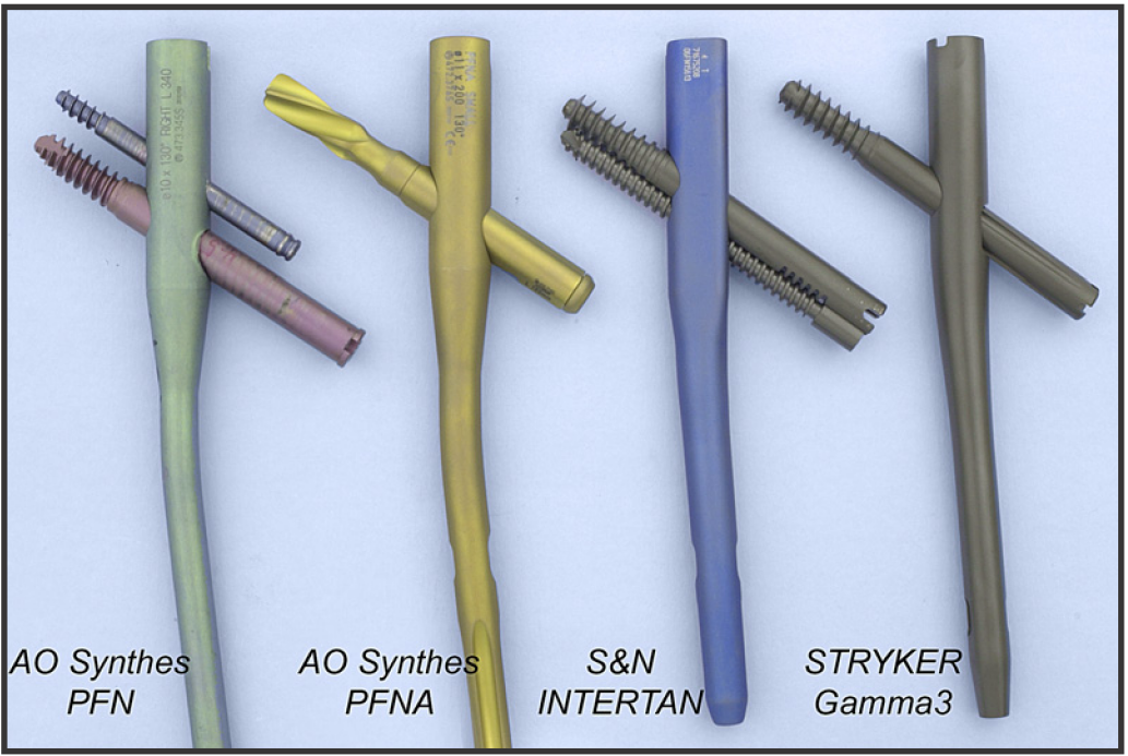
IV. Implant Selection and Failure Modes: PFNA vs. InterTAN
This is also a classic controversy. According to recent meta-analyses and biomechanical studies, each has advantages and disadvantages; selection should be individualized.
1. Cut-out vs. Cut-through
These are two different concepts and must be clearly distinguished.
-
The Achilles heel of PFNA (helical blade)—cut-through (medial penetration):
- Mechanism:The helical blade provides good rotational stability, but because it lacks threads, its resistance to axial sliding is weak. In patients with severe osteoporosis, if varus collapse occurs, the blade does not slide out laterally; instead, the femoral head “swallows” the blade, causing medial cut-through into the acetabulum.
- Countermeasure:In severe osteoporosis, be cautious with blade systems, or use cement augmentation.
-
Advantages of InterTAN (integrated interlocking nail):
- Mechanism:The dual screws form a “box” construct, providing stronger rotational stability; threaded engagement resists axial sliding, with a very low risk of cut-through.
- Fracture-prevention design:The distal clothespin split of InterTAN reduces stress concentration and lowers the risk of distal femoral shaft fracture.
2. How to Choose?
- PFNA/TFNA:Easy for even novice surgeons, shorter operative time, and less blood loss. Suitable for extremely frail elderly patients for whom this is essentially a life-saving/damage-control procedure.
- InterTAN: More complex surgical steps and slightly more blood loss. However, it provides better stability and stronger resistance to rotation and shortening. Suitable for unstable fractures (31-A3), relatively good bone quality, longer life expectancy, and patients with higher functional demands.
V. Key Metrics: TAD and CalTAD
Regardless of which nail is used, the final step must not be wrong.
- TAD (Tip-Apex Distance):Must be <25 mm.
- CalTAD (calcar–tip apex distance):This is a more sensitive metric. On the AP view, the screw should be close to the calcar (inferior cortex), and on the lateral view it should be centered.
- Positioning mnemonic: Inferior on AP, center on lateral. Avoid superior placement—that is the “death zone.”
Summary
The key to completing this procedure and avoiding pitfalls is not how expensive the nail is, but:
- Never accept varus reduction (mini-open if necessary).
- Respect anatomy(note the anterior bow ROC; use a blocking pin if necessary).
- Precise screw placement (TAD <25 mm; follow the calcar on AP view).
- Individualize implant selection (prevent cut-through in severe osteoporosis; choose InterTAN for unstable fractures).